
Biomimicry is considered the bridge between biology and design, in which researchers observe traits in nature and adapt these natural models for human technology, systems, and design. These nature-inspired innovations are as wide-ranging as patterning the desalination of soil on the vascular systems of plants; mimicking the hooks on burrs for the development of Velcro®, creating flexible robots based on the structure of snake skeletons; and designing fans and propellers with inspiration from the nautilus shell’s beautiful interior spiral staircase (Figure 1).
Micro-CT offers researchers an ideal non-destructive approach to internal and external views of natural structures and materials, at macro-, micro-, and nano-scale. The detail provided by micro-CT allows scientists to basically reverse-engineer nature in 3D. These biometric studies are applied across many applications, including the investigation of multifunctional and hierarchical structures and the growing use of additive manufacturing.
This month we look at various micro-CT studies that demonstrate the unique synergy between micro-CT and biomimicry.
X-ray Microscopic Examination of an Apple
Salt-contaminated soil requires treatment to eliminate high levels of sodium chloride created by soil salinization in agriculture or in saltwater spills in the oil and gas industry. The biometric process for desalination of soil mimics the way plants transport water and salt by using evapo-transpiration to move saltwater above the soil surface, where it can be treated.
High-resolution X-ray microscopy is increasingly used for structural phenotyping studies for plant biology at macro-, micro-, and nanoscale levels. To demonstrate the benefits that arise from having one instrument with multiple detectors, we imaged an apple at three different scales using our Bruker SkyScan 2214 Multi-scale X-ray Nano-CT. Micro-CT and nanotomography are now used to study water transport in plants; for 3D imaging of plant tissues and organs; and for the study of plant development and organ morphogenesis, non-destructively and with minimal or no sample preparation.
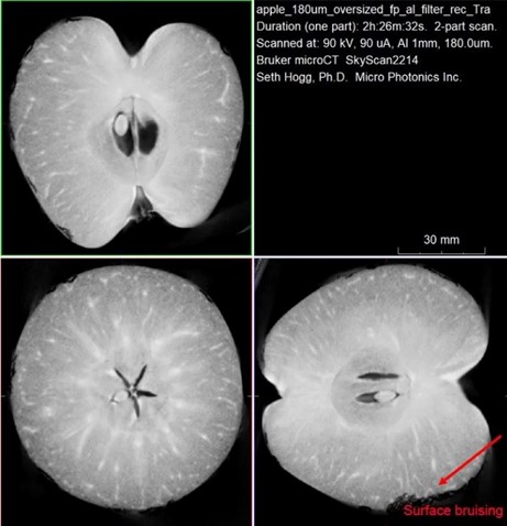
As shown in Figure 2, the overview imaging of the apple provided by the flat panel detector allows us to view hidden interior details such as fruit bruising as well as the location of the core, seeds, and the intricate vascular network responsible for transporting water and sugars through the xylem and phloem.
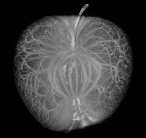
Since the vasculature network within the fruit contains concentrated sugars and other transported molecules, we observe natural contrast from the rest of the fruit. Utilizing a maximum intensity projection (MIP) view within CTVox provides us with an interactive 3D view of the complex paths mapped out by the vasculature (Figure 3).
X-ray Microscopic Examination of a Cocklebur (Xanthium)
Nature has developed many unique structures that scientists use for inspiration in developing innovative technologies through biomimicry. Velcro was invented by George de Mestral in 1941 and was inspired by the burrs he found on himself and on his dog. Being an engineer and entrepreneur, he realized the small hooks of the burr and loops of the fur/fabric allowed the burr to adhere exceedingly well. This sparked his idea to mimic the structure as a potential fastener.
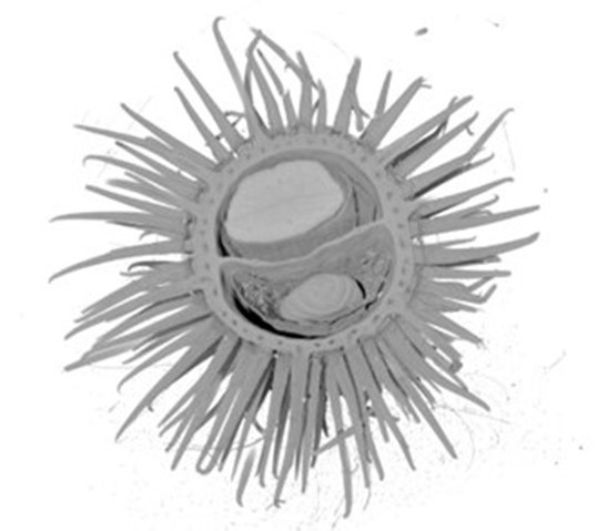
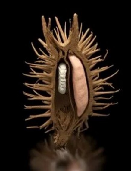
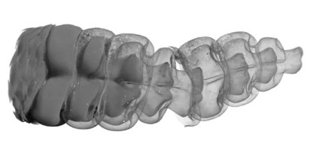
We examined a cocklebur sample removed from a dog’s fur and found an unexpected surprise using our high-speed SkyScan 1275 micro-CT at an isotropic voxel size of 10µm.

As shown in Figure 4, the SkyScan 1275 micro-CT resolved the large structures of the cocklebur sample and provided us a high-resolution view of the interior contents of the burr. While burrs are used in nature to spread seeds and propagate a plant’s survival in the ecosystem, in this case it appears a parasitic insect species was also present in our sample. In looking at the chambers within the cocklebur, we see the large seed present in one of the two central chambers. However, within the other chamber the seed is missing and is instead replaced by a small pupa, which likely consumed the seed during its growth. The bright white spots within the image are dense inclusions present within the pupa itself and could represent locally high concentrations of either inorganic components or even metals.
X-ray Microscopic Examination of a Rattlesnake
Snakes are elongated, limbless, carnivorous reptiles with a uniquely flexible body that can maneuver in small spaces by bending their spines into serpentine coils or using their scales to push off objects. They provide a biological model for robots designed for remote navigation and the inspection of small, otherwise inaccessible spaces that are dangerous or difficult for humans. Like snakes, these robots can operate in mud, water, and sand, and can manage through irregular surfaces, such as debris.
We imaged a rattlesnake at three scales using the SkyScan 1273 micro-CT. The large sample volume of the SkyScan 1273 allowed us to examine the snake at a full body overview scale (Figure 7) and two higher resolution captures of regions of high interest – the tail (Figure 8) and head (Figures 9).
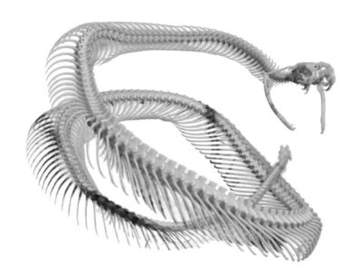
In examining the head, the bone structure shows great detail in the jaws and teeth with multiple rows of teeth visible within the head (Figure 9). The unique feature of many snakes, where the bottom jaw halves are only connected by soft tissue allowing for the consumption of large prey, is also evident.

X-ray Microscopic Examination of a Nautilus Shell
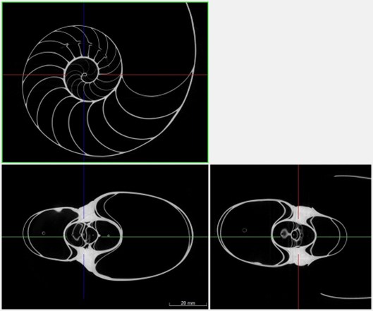
Within the beauty of the chambered nautilus is a remarkable symmetry created by the concentric rings within the shell. This distinctive pattern is often related to the Fibonacci Sequence, also known as the Golden Number, although the shells do not exactly match the mandatory proportions for this numerical pattern.

Aside from its beauty, the nautilus shell has inspired designs for fans and propellers that will move air or fluids with the least amount of effort. The soft-bodied nautilus or cephalopod (a type of mollusk) lives inside its hard external shell, and it creates the interior of the shell for the best path outside for food, and then to return inside safely. The spiral shape thus created provides the least resistance to water or air passing through it. Cross sections of that spiral shape (Figure 11) are inspiring researchers to redesign powered devices that push, pull, or mix fluids, such as water and sewer pumps and macerators; and coolant systems in engines and even nuclear power plants.
Using nature’s method of streamlining with the spiral shape, engineers are creating highly efficient fans and propellers, which look like regular fan blades but which optimize how air and water move along the blade.
Conclusion
We hope you found this Image of the Month informative and encourage you to subscribe to our newsletter and social media channels in preparation for the continuation of our Image of the Month series next month.
These scans were completed on our Bruker SkyScan systems at the Micro Photonics Imaging Laboratory in Allentown, PA. For scan specifications or details on reconstructions, visualizations, and volumetric inspection of the 2D and 3D results, please follow the links below:
Would you like your work to be featured in our monthly newsletter? If so, please contact us by calling Seth Hogg at 610-366-7103 or e-mailing seth.hogg@microphotonics.com.