The Symmetry of Mathematics and Nature: Using Micro-CT to Examine a Nautilus Shell
Within the beauty of the chambered nautilus is a remarkable symmetry created by the concentric rings within the shell. This distinctive pattern is often related to the Fibonacci Sequence, also known as the Golden Number, although the shells do not exactly match the mandatory proportions for this numerical pattern1: 1, 1, 2, 3, 5, 8 … with the remainder of the numbers corresponding to the sum of the two numbers proceeding them in the sequence2.
The chambered nautilus is commonly referred to as a living fossil as it has undergone little change in more than 400 million years3. Grouped along with squid, cuttlefish, and octopus, the nautilus is a member of the cephalopod family and holds the distinction of being the only family member with a fully developed shell. Perhaps this helps to compensate for the undeveloped eyes leading to poor vision similar to a pinhole camera3. Born with about four chambers, the nautilus will eventually grow to an average of 30 chambers by maturity. Despite having so many chambers, the animal only resides in the outermost chamber while the remainder of the chambers are used to control buyoyancy3.
Micro-CT is a powerful tool for examining finely detailed structures such as the nautilus since we can see inside the sample without physically cutting the shell in half to view the chambers. The resulting three-dimensional reconstruction is captured with a high resolution, allowing the end user to digitally dissect the sample in any orientation. In this situation, we chose to utilize our SkyScan 1173 system because of its large sample capacity and fine detail resolution.

Micro-CT Scan of Nautilus Shell
After reconstructing the X-ray attenuation data into representations of the three-dimensional structure we can observe the different chambers within the internal structure of this shell from different planar views (Figure 1).

The spiral geometry of the chambers is even present when examining the two dimensional projection images, which comprise the raw results from this scan (Figure 2).
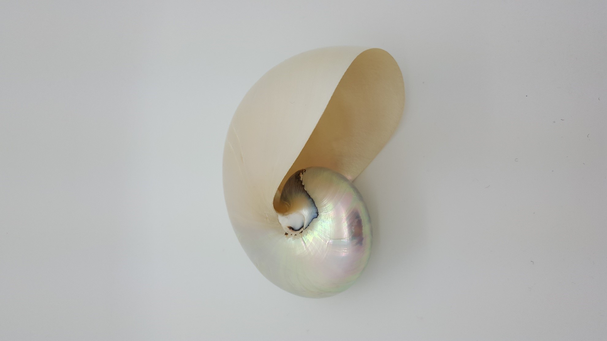
Using micro-CT to examine the nautilus shell demonstrates the power derived from having the ability to inspect the internal three-dimensional morphology of samples. Looking at the outside of this sample alone it would be hard to imagine all the intricate details contained within (Figure 3).
Conclusion
Micro-CT excels as a tool to nondestructively examine the internal structure of animal samples and produces a highly detailed copy suitable for digital dissection. We hope you found this image of the month interesting. If you have an image of the month sample that you would like us to scan, please contact us by calling Seth Hogg at 610-366-7103 or e-mailing seth.hogg@microphotonics.com
Scan Specifications
| Sample | Nautilus |
| Voltage (kV) | 130 |
| Current (µA) | 60 |
| Pixel Size (µm) | 55 |
| Rotation Step | 0.4 |
| Scan Time (HH:MM:SS) | 02:27:26 |
All scans completed on our high-speed SkyScan 1173 micro-CT system at the Micro Photonics Imaging Laboratory in Allentown, PA. Reconstructions were completed using NRecon and visualization of 2D and 3D results were completed using DataViewer and CTVox.
Works Cited
- https://www.goldennumber.net/nautilus-spiral-golden-ratio/
- http://mathforum.org/mathimages/index.php/Fibonacci_Numbers
- https://aqua.org/explore/animals/chambered-nautilus