Audrey Williams, MS
Missouri University of Science and Technology
Jerald D. Harris, PhD
Professor Emeritus (Earth and Environmental Sciences)
Utah Tech University
This Researcher Spotlight highlights the work of Audrey Williams, who used micro-CT to non-destructively produce images that resembled traditional, physical, histological thin sections, preserving the fossil bone. Her advisor and co-author, Dr. Jerry Harris, is a vertebrate paleontologist recognized for his extensive work on dinosaurs and prehistoric tracks. Micro-CT imaging and subsequent image processing were performed by Seth Hogg, PhD, at the Micro Photonics laboratory in Allentown, PA.
Sauropod dinosaurs, the largest terrestrial animals, have long been studied to understand how their anatomy supported their massive bodies—particularly their elongated necks. However, the absence of clear modern analogs and the poor preservation of soft tissues have limited direct evidence of their neck support structures. This study investigates the nature of sauropod neck ligaments using histology, micro-CT imaging, and comparative morphology. The results suggest that, rather than a mammal-like nuchal ligament, sauropods likely possessed dorsally positioned supraspinal ligaments and possibly interlaminar elastic ligaments. These findings provide new insights into sauropod neck support, with implications for their mobility, feeding behavior, and biomechanics.
Can you expand briefly on your research focus?
I was interested in finding out what ligaments two of the giant, long-necked sauropod dinosaurs, Apatosaurus and Diplodocus (both of which belong to the diplodocid group of sauropods), had for holding their necks up constantly. It likely wasn’t just muscles all the time—imagine holding your arm straight out to the side for hours on end, how tired your arm muscles would get working against gravity for so long! So, these sauropods probably had some ligaments among their neck vertebrae that held their necks up without needing much help from muscles. The previous schools of thought had three different ligament types that have been debated over the past few decades that would have resulted in different neck orientations: either more vertical or more horizontal. Most recently, it was proposed that diplodocid sauropods had more horizontal neck orientations throughout their lives, meaning that, like having your arm straight out to the side, it would burn a lot of calories firing the muscles to hold all that weight up without help from ligaments. But because ligaments generally don’t preserve as fossils, I had to look for signs on the preserved neck vertebrae that indicated what ligaments were present that could have helped hold the neck up while expending minimal energy.
My advisor, Dr. Jerry Harris, suggested histological examination—looking at microscopic, interior features of bone by slicing the bone (physically or digitally) into thin sections. I made thin sections from the bone at the tips of the tall spines (called neural spines) that stick off the tops of the neck vertebrae, where ligaments would likely attach, of Apatosaurus and Diplodocus and examined them under a microscope. I did this to see the orientation of bone fibers at the point of likely attachment and used that to infer the type of ligament system acting on those spines. I also incorporated micro-CT imaging to examine the bones non-destructively. The results of my research, through physical thin section preparation and the micro-CT images from Micro Photonics, were some of the first images showing orientation that support the more-horizontal-neck hypothesis that paleontologists have been discussing for diplodocid sauropods.
What is important about this research today, and what are you hoping to achieve through your work?
This research provides a means of inferring the natures of soft-tissue structures that were present in extinct species, even when those soft tissues weren’t preserved for study. I was hoping to find evidence of specific types of bone orientation that could specify a specific ligament system at a micro-level examination, instead of making broad-level hypotheses, as had been used primarily in the past, but not specifically tested. This preliminary research has exceeded my expectations by exposing crystal-clear orientations that can easily be seen with the naked eye. Similar work could be done on any extinct animals to see what kinds of ligament systems they employed, too. The work is very broadly applicable!
How are you using micro-CT technology in your research?
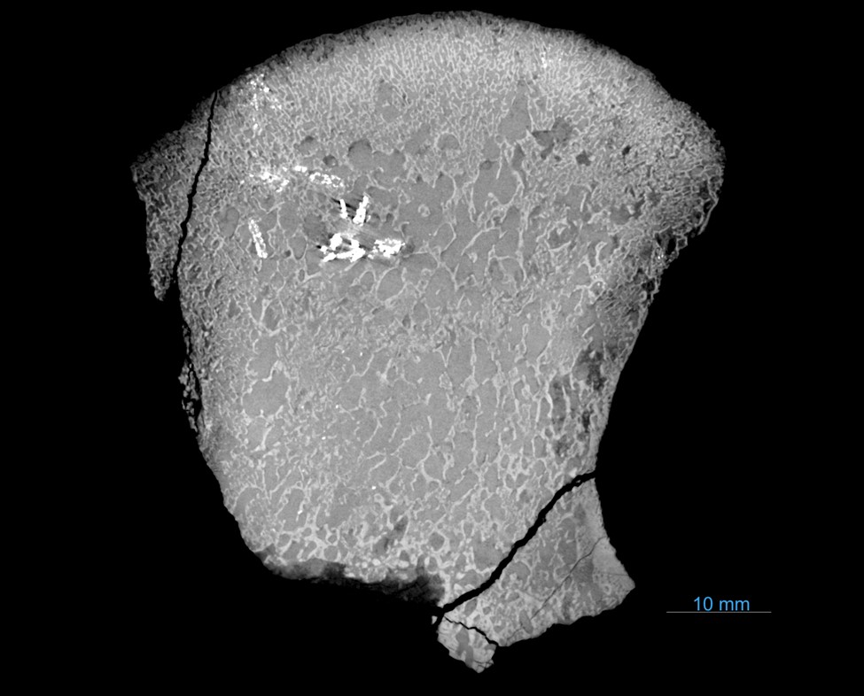
The unfortunate reality of making traditional, physical, thin sections from fossil material is that once the bone is cut, it is forever damaged. I studied the abilities of micro-CT instruments and how they can produce detailed digital images of fossil bone. I wanted to see if micro-CT scanning could produce digital thin sections that could not only show the orientations of the internal bone structure but, more importantly, find a way to prevent unnecessary destructive testing. The images from Micro Photonics exceeded my expectations: the instrument produced raw images that showed the detailed network of the cortical (dense) and trabecular (spongy) bone. With the use of the 3D software Dragonfly, the images were further enhanced and provided clear images that resembled traditional, physical, histological thin sections.
Are there any images that you could share illustrating results using micro-CT with your application?
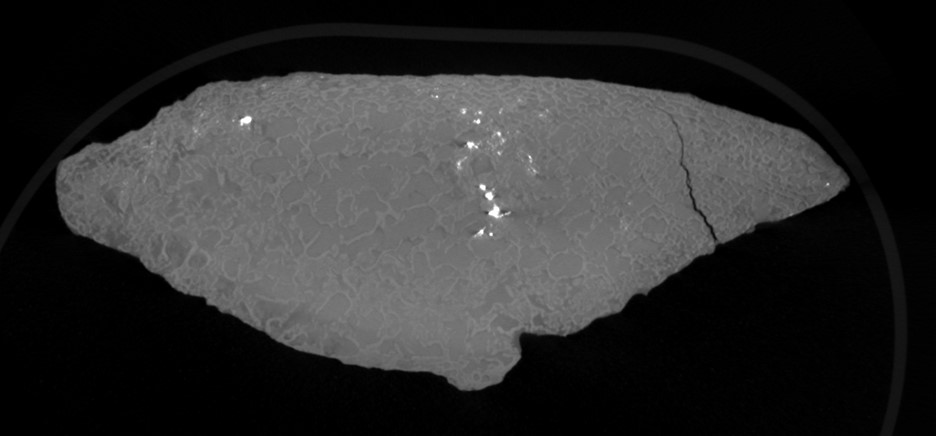
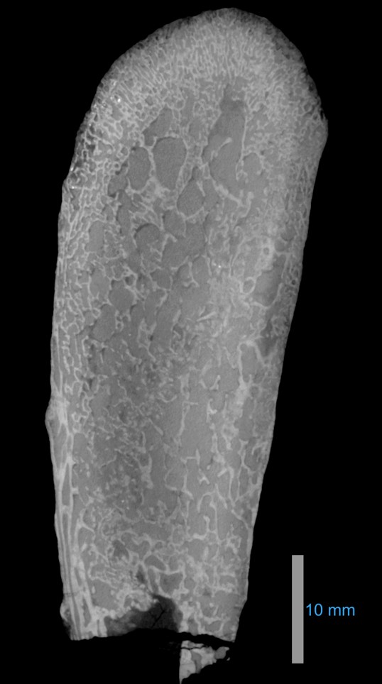
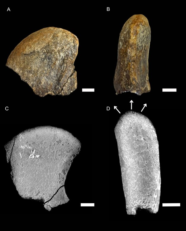
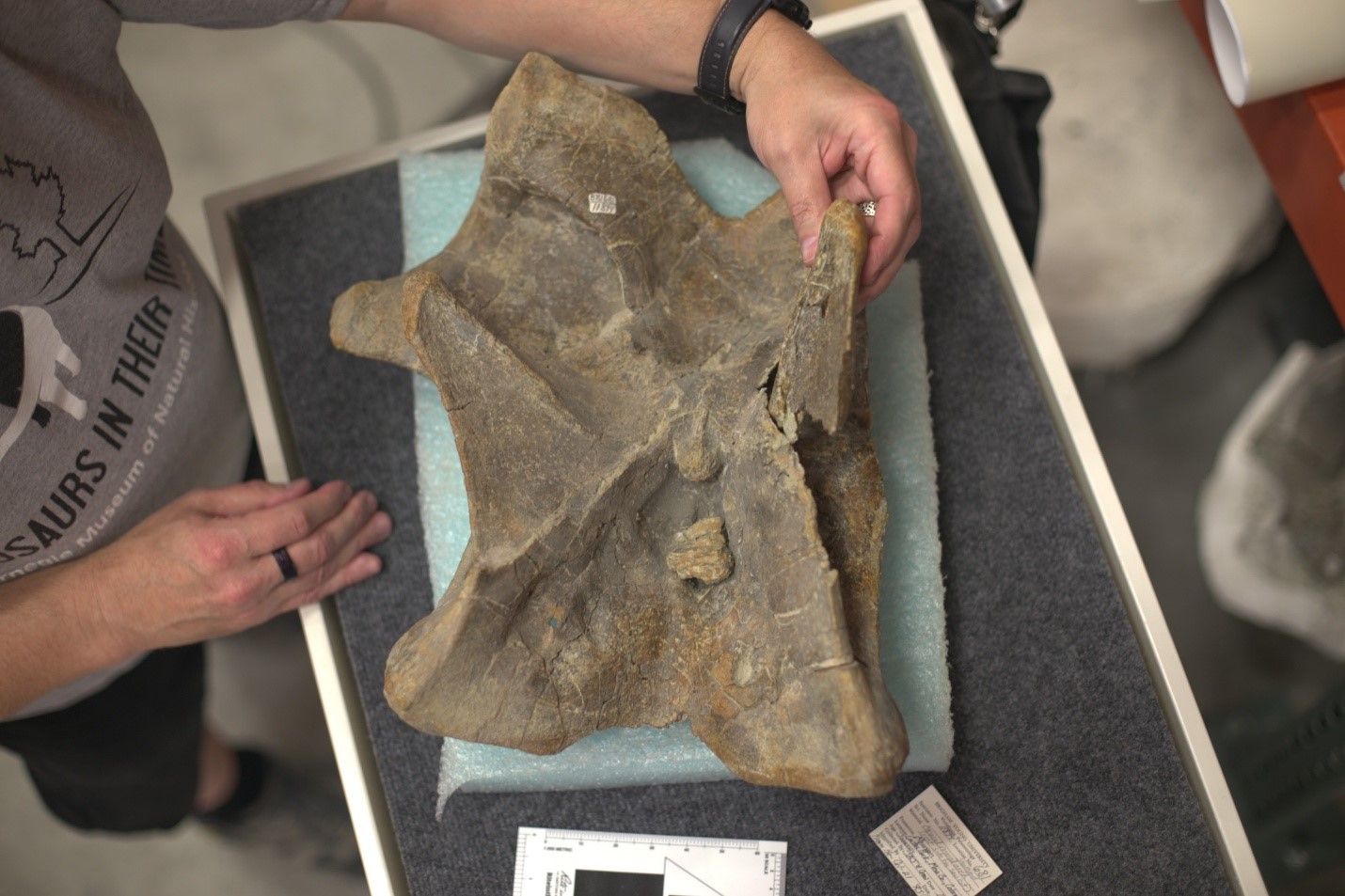
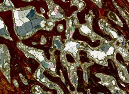
Figure 7: An image of the physical, thin histological sections taken from the tops of the neck vertebrae.
What has been the most important discovery or unexpected outcome in your research?
I began my research by looking for evidence of a nuchal ligament, a strong, rubbery neck ligament that is commonly observed in mammals with heavy heads and long necks, such as giraffes, bison, and horses. This type of ligament runs from tall spines in the shoulder region to the back end of the skull, and it works similarly to how cables in a suspension bridge work: the ligament has a strong cord above the levels of the neck vertebrae, and extensions from that cord down to each vertebra—the neck is literally suspended from this ligament, holding the neck up. Unexpectedly, my research indicated a ligament system more like that of reptiles, with a supraspinal ligament instead. This type of ligament is a flatter cord that runs directly from the tip of one neck vertebra’s spine to the next, all the way from the back to the skull. Although it didn’t involve thin sections or micro-CT, I also found suggestive, but as-yet untested, evidence that Apatosaurus and Diplodocus also had a bird-like system of additional neck ligaments, called interlaminar elastic ligaments, that would also have aided in holding up their necks, which was also novel.
Key publications for further reading:
Williams, Audrey R. and Harris, J.D. (2025) Cervical ligament systems in sauropod dinosaurs: what support is there? Vertebrate Anatomy Morphology Palaeontology, 13, 81–97. https://doi.org/10.18435/vamp29412
Harris, J.D. (2024). What exactly is a nuchal ligament and who exactly has one? Vertebrate Anatomy Morphology Palaeontology, 12(1): 59–80. https://doi.org/10.18435/vamp29405