
Micro-CT-based morphological parameters are often used for assessment of 3D bone morphology in studies of small animals. Ex-vivo micro-CT imaging of mouse bones provides a rapid and reliably proving way for measuring many different morphological parameters that make suitable markers for bone growth and general health. These traits are often explored in larger studies to help understand differences between among treatment groups in a research study.
SkyScan desktop ex-vivo instruments are a popular choice in Research and industrial settings after their introduction to the field more than fifteen years ago, and several of the SkyScan instruments have excellent capabilities for most bone imaging projects.
X-Ray Microscopic Imaging of a Mouse Femur
We examined a dissected mouse femur using each of the desktop ex-vivo micro-CT instruments in our office for comparison. We kept the same isotropic voxel size of eight micrometers but used suggested image capture settings specific to each instrument as recommended in the associated method notes. We imaged the same bone mineral density (BMD) calibration phantoms with each sample to allow us to compare the quantification of tissue mineral density (TMD) across each instrument as well.

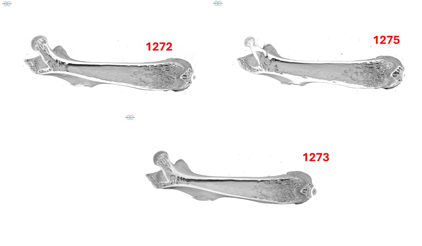
As shown in Figure 2, DataViewer allows us to examine each dataset in a pseudo 3D viewing mode. While each dataset was collected using slightly different settings specific to each instrument, the overall quality of each dataset is fairly similar. All the available SkyScan instruments are a great match for this project type and thus, when deciding which system to use, we often consider other factors such as whether throughput or resolution are the most important variables to optimize against.
For this project, both the SkyScan 1272 and SkyScan 1275 were able to capture the full bone volume with an imaging time of under an hour with the SkyScan 1275 being almost twice as quick as the SkyScan 1272. This makes sense because the scintillator design of the SkyScan 1275 favors acquisition speed (150ms exposure) over resolution, unlike the SkyScan 1272 that favors resolution over acquisition speed (645ms exposure).
The SkyScan 1273 took a bit longer than both other systems as it was operating within its small spot size mode to better capture details within the sample. However, it provides the most versatility of the desktop instruments as it scales well from small all the way through large samples (~250mm) and is optimized to be a balance between resolution and imaging speed for most samples.
As we observed in DataViewer, a comparison of the samples within CTVox shows limited differences in image quality among the datasets, as shown in Figure 3. Each dataset provides a good balance of resolving thick cortical bone while also capturing thin trabecular structures.
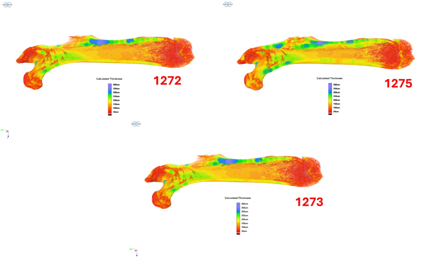
After running each sample through CTAnalyzer (CTAn), we extracted quantitative data for the sample from each instrument, including the localized thickness throughout the bone, the average bone thickness, and the TMD. Figure 4highlights the localized thickness datasets corresponding to each instrument. For each dataset, the same task list was used with minor differences in the segmentation values to account for differences in the grayscale values in each reconstructed dataset. In all cases, we attempted to utilize segmentation values that would achieve a similar selection for each sample. To calculate TMD, each instrument also imaged a calibrated pair of phantoms, as highlighted in Bruker Method Note 9.
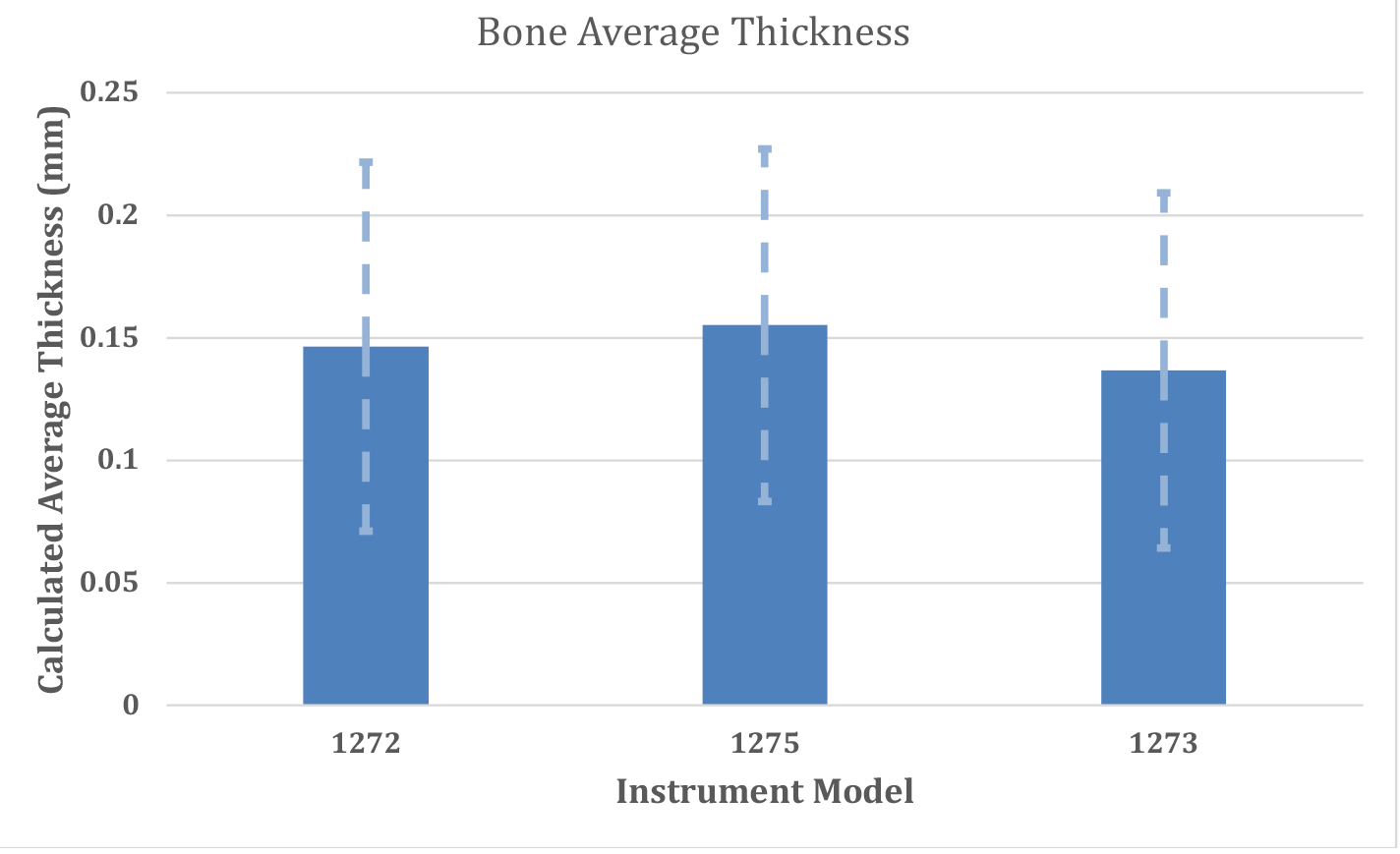
Figure 5 provides a numerical context to the color plots presented in the previous figure and demonstrates the high degree of agreement among the three instruments for the imaged femur. While minor differences exist for the average thickness as calculated from each instrument, the scale of the differences is small in comparison to the standard deviation of the values themselves arising from the wide distribution of thickness in the whole bone sample.
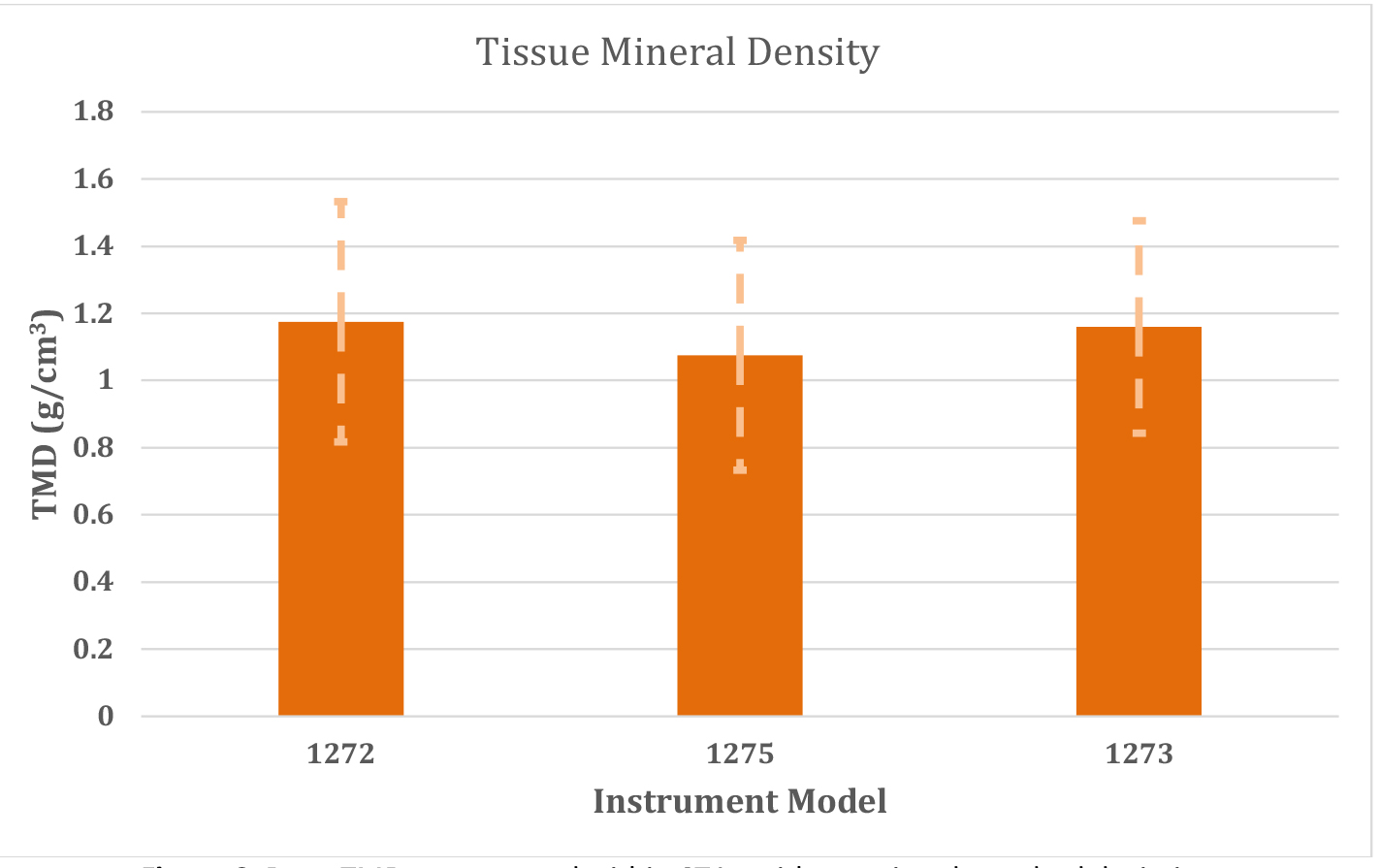
Figure 6 presents similar quantitative data for each instrument in calculating the whole bone density, known as the TMD. As we observed for thickness, minor differences between each instrument exist for the calculated density, but the magnitude of the differences is far below the associated standard deviation with each calculated TMD value.
Conclusion
The SkyScan product line represents a large set of capabilities for most bone imaging projects and has been a key player in this area for many years. While each of our ex-vivo systems have unique features and considerations, they all are a great choice for mouse bone imaging. Though some minor differences exist in the quantitative output for each instrument, the differences among the datasets were minimal.
We hope you found this Image of the Month informative and encourage you to subscribe to our newsletter and social media channels in preparation for the continuation of our Image of the Month series next month.
Scan Specifications
| Instrument | 1272 | 1275 | 1273 |
| Voltage (kV) | 70 | 80 | 70 |
| Current (µA) | 142 | 125 | 114 |
| Filter | 0.5 mm Aluminum | 0.5 mm Aluminum | 0.5 mm Aluminum |
| Voxel Size (µm) | 8 | 8 | 8 |
| Rotation Step | 0.4 | 0.4 | 0.3 |
| Exposure Time (ms) | 645 | 150 | 216 |
| Rotation Extent (deg.) | 180 | 180 | 180 |
| Scan Time (HH:MM:SS) | 00:58:14 | 00:44:24 | 02:17:46 |
These scans were completed on our SkyScan 1272, 1275, and 1273 micro-CT systems at the Micro Photonics Imaging Laboratory in Allentown, PA. Reconstructions were completed using NRecon 2.0 while visualization and volumetric inspection of the 2D and 3D results were completed using DataViewer and CTVox. A bone dataset was also converted to a STL volumetric model using Synopsys’ Simpleware ScanIP software with the CAD add-on module (Synopsys, Inc., Mountain View, USA) before 3D rendering using Maverick Render Indie (Random Control, Madrid, Spain.
Would you like your work to be featured in our monthly newsletter? If so, please contact us by calling Seth Hogg at 610-366-7103 or emailing seth.hogg@microphotonics.com.
*Simpleware software (Synopsys, Inc., Mountain View, USA) enables you to comprehensively process 3D image data (MRI, CT, micro-CT, FIB-SEM…) and export models suitable for CAD, CAE and 3D printing. Use Simpleware software’s capabilities to visualize, analyze, and quantify your data, and to export models for design and simulation workflows. Simpleware™ is a trademark of Synopsys, Inc. in the U.S. and/or other countries.