
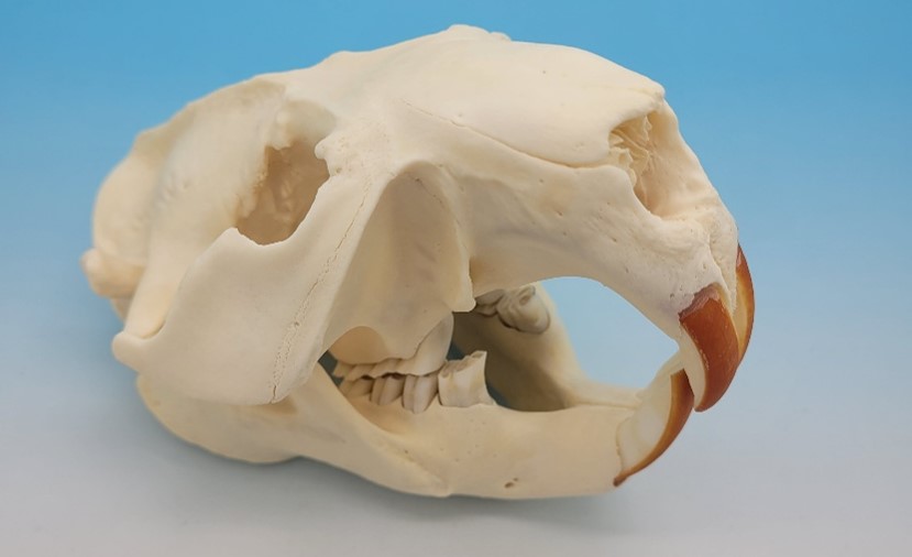
The micro-CT images in this study of an American beaver skull highlight the ability to examine fine structural details in a specimen far larger than the mice and rats we typically image. Using the large imaging volume of the SKYSCAN 1273, we acquired both standard and high-speed datasets of the skull while maintaining high image quality across the entire specimen.
Micro-CT imaging of a large object such as a beaver skull presents a significant technical challenge: achieving high-resolution detail over a large field of view. In this study, we demonstrate that a high-resolution dataset (50 µm isotropic voxel) provides an effective balance between visualizing delicate inner skull structures and penetrating the dense enamel coating of the molars.
The beaver’s prominent, ever-growing incisors are among its most striking features. These teeth owe their reddish-orange coloration to ferritins—iron-containing proteins embedded within the enamel between granules of calcium hydroxyapatite.¹ Compared with typical rodent bone, teeth present greater thickness and mineral density, requiring an appropriate micro-CT solution. The SkyScan 1273 readily images both small and large teeth, including fossilized specimens affected by mineral diagenesis.
X-Ray Microscopic Imaging – High Resolution Imaging of Biological Specimens
Micro-CT is an ideal tool for imaging bone and teeth, revealing fragile internal anatomy non-destructively and in high detail. For this study, the beaver skull dataset was acquired using our high-resolution SkyScan 1273 at isotropic voxel sizes of 50 and 100 µm, providing us with a high-resolution view into fine skull structures and teeth. With the ability to image a field of view of up to 10 inches in height and width, the SkyScan 1273 provides the most flexibility on sample size of any instrument in our portfolio. The SkyScan 1273 also offers a unique, high-speed continuous imaging mode, which provides an approximate 90% time saving with a tradeoff for a 50% reduction in resolution. This versatility makes the SkyScan 1273 a popular choice for many laboratories around the country that desire to image the broadest collection of specimens possible in applications such as comparative zoology.
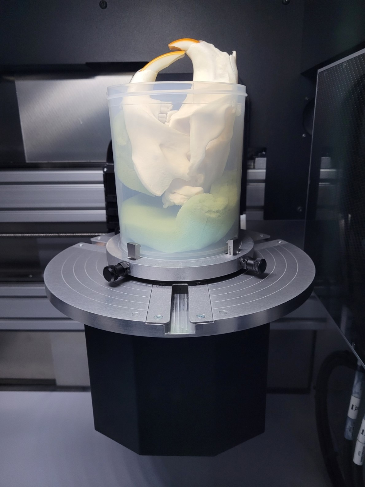
As shown in Figure 2, we used the large imaging volume within the benchtop SkyScan 1273 instrument by mounting the beaver skull in a large plastic jar with low-density upholstery foam selectively placed to ensure the sample remained stationary during imaging. Due to the density of bone and teeth, the signal in the dataset arising from the foam blocks is negligible and does not impact our final visualization of the results.
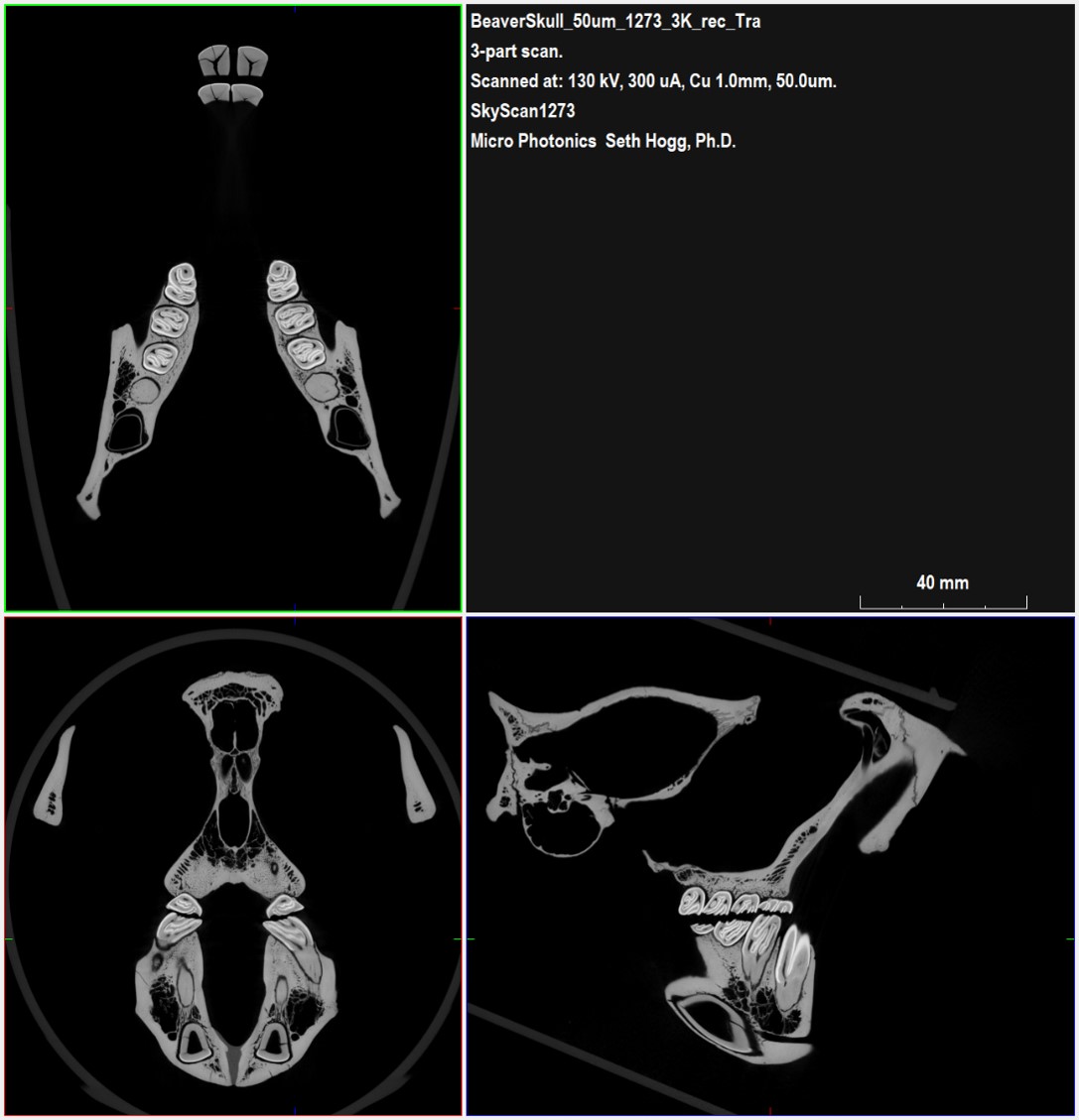
As shown from Bruker DataViewer in Figure 3, our high-resolution (50 µm isotropic voxel) dataset balances the visualization of both thin bone structures in the inner skull along with the dense enamel coating present on the animal’s molars.
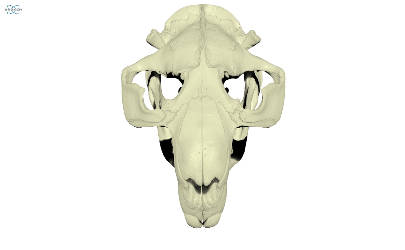
For this project, we utilized Bruker CTVox to render the volumetric data into an interactive dataset and export videos exploring the skull’s structures. Figure 4 provides a face-on view from CTVox showing the front of the skull, including the eye sockets and nasal passages.
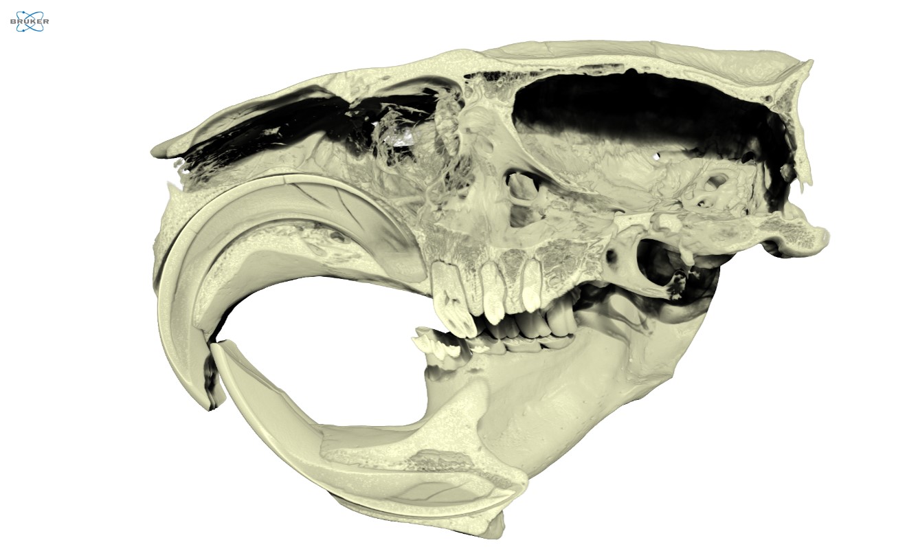
CTVox also allows us to control sliced planes through the dataset, enabling us to visualize and study internal features and how components are connected within samples. Figure 5 highlights a clipped view along a set of molars in the jaw, letting us qualitatively observe the enamel and dentin along with the alveolar bone surrounding each tooth.
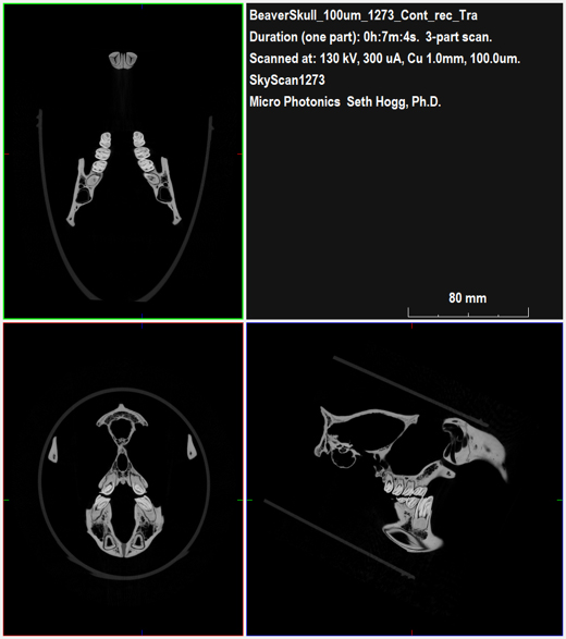
Figure 6 provides a rendered image of the second beaver skull datasets acquired using the continuous imaging mode, allowing for comparison to the earlier images (Figure 3). While some fine detail is lost through the doubling of our voxel size, the switch to continuous imaging reduces the total dataset acquisition time from over nine hours to only about twenty minutes while still retaining many key details. For projects where the overall goal is general exploration or for cataloguing collections, the throughput available with the reduced imaging times more than justifies the slight cost in overall resolution.
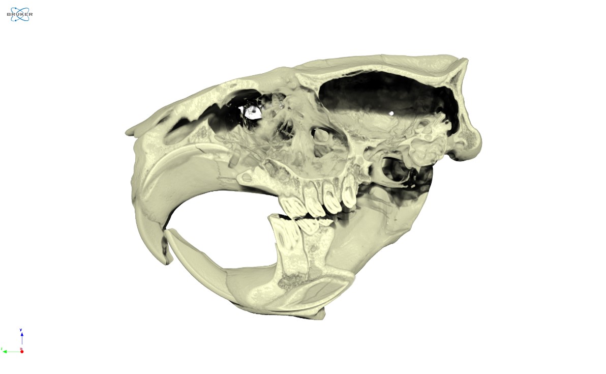
Figure 7 also provides a clipped view with CTVox in direct comparison to the higher-resolution dataset (Figure 5). As we observed for the planar slices, while the resolution of the continuous imaging dataset is reduced, the overall level of features captured remains excellent including even a crack visible in a lower incisor.
Conclusion
Micro-CT is a versatile technique allowing the SkyScan 1273 to image almost any sample type that both fits within the instrument and allows for X-ray energy to pass through without full attenuation. In this case, we were able to fit an adult American Beaver Skull comfortably within the SkyScan 1273 and acquire two different datasets. Each dataset allowed for the visualization of both thin, structural bone within the skull as well as fine details related to each tooth.
We hope you find this Image of the Month article informative and encourage you to subscribe to our newsletter and social media channels in preparation for the continuation of our Image of the Month series next month.
Scan Specifications
| Imaging Mode | Step-and-Shoot | Continuous |
| Voltage (kV) | 130 | 130 |
| Current (µA) | 300 | 300 |
| Filter | 1 mm Copper | 1 mm Copper |
| Voxel Size (µm) | 50 | 100 |
| Rotation Step (deg) | 0.25 | Continuous |
| Exposure Time (ms) | 692 | 692 |
| Rotation Extent (deg.) | 360 | 360 |
| Scan Time (HH:MM:SS) | 09:20:51 | 00:21:12 |
These scans were completed on our Bruker SkyScan 1273 instrument at the Micro Photonics Imaging Laboratory in Allentown, PA. Reconstructions were completed using NRecon 2.0 while visualization and volumetric inspection of the 2D and 3D results were completed using DataViewer and CTVox.
Would you like your work to be featured in our monthly newsletter? If so, please contact us by calling Seth Hogg at 610-366-7103 or emailing seth.hogg@microphotonics.com.
References