
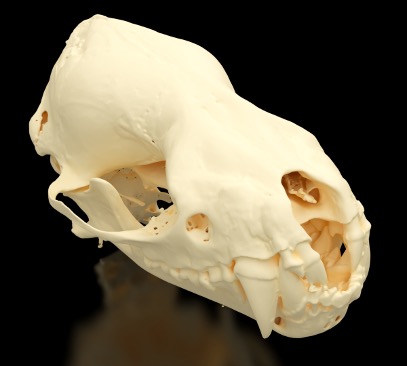
Bats account for more than one in five living mammal species, representing an extraordinary diversity of feeding strategies, sensory adaptations, and ecological specializations1. From bats that navigate in complete darkness to those that feed exclusively on blood, the extraordinary diversity of bat behavior is reflected in the specialized anatomy of their skulls.
Bat skulls provide valuable insights into the relationships between anatomy, function, and evolutionary adaptation. With more than 1,400 species occupying diverse ecological niches, bats exhibit remarkable variation in cranial morphology associated with echolocation, hearing, feeding behavior, and sensory specialization. Differences in skull shape, jaw structure, dentition, nasal cavities, and the bony structures of the inner ear reflect adaptations to specialized diets and methods of navigation. High-resolution micro-CT enables researchers to examine these complex anatomical features in 3D, including delicate internal structures that are difficult to visualize using conventional imaging or physical dissection. Quantitative analysis of cranial geometry, bone morphology, and internal cavities supports comparative studies of evolutionary relationships, functional biomechanics, and species-specific adaptations. Importantly, the non-destructive nature of micro-CT also makes it particularly valuable for examining rare, fragile, and irreplaceable museum specimens.
This study highlights the SkyScan 1273’s significant capabilities for comparative biology and for small animal imaging. With the ability to image samples ranging from a few millimeters in size up to about 25 centimeters, the SkyScan 1273 is a versatile instrument that provides the broadest range of imaging capabilities within our current portfolio. For this study, a bat skull was examined with limited sample preparation. The SkyScan 1273 successfully resolved the fine bony structures present throughout the skull, allowing for further examination by researchers.
Micro-CT Imaging of Bones
The bat was imaged in its naturally deceased state, with minimal sample preparation required. The specimen was carefully secured within a small polymer tube to ensure stability during scanning. Using the SkyScan 1273’s flat-panel detector, fine anatomical details were captured at an isotropic voxel size of 15 µm, enabling high-resolution, 3D visualization of the skull’s delicate bony structures.
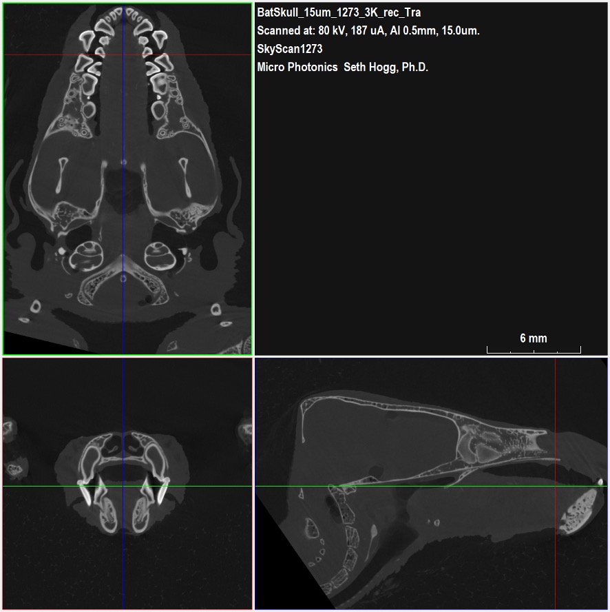
As shown in Figure 2, fine trabecular bone structures are present throughout the skull and are well resolved by the isotropic 15 micrometer voxel size used for this study. We also observe the local brightening in the dataset arising from the enamel present on the teeth due to the higher attenuation of X-ray energy by the enamel compared to the dentin and the surrounding bone structures.
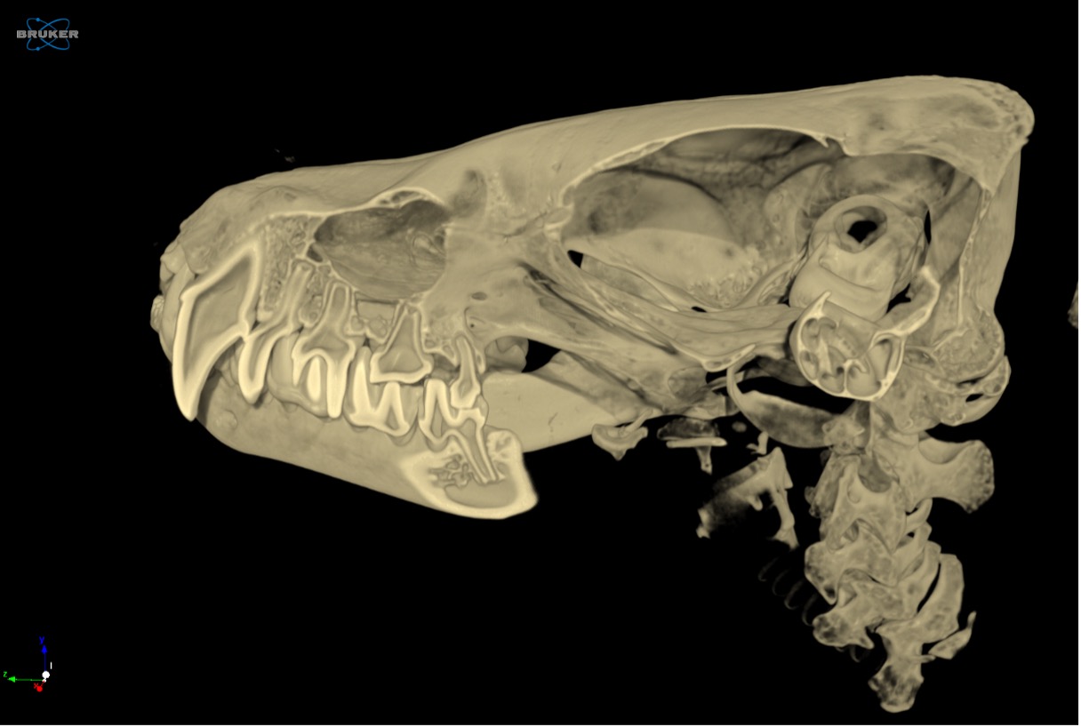
For this project, we utilized Bruker CTVox to render the volumetric data into an interactive dataset to further explore fine bone and teeth structures present within the bat skull. Figure 3 provides a clipped view from within CTVox looking along the length of the skull with the interface between the teeth and the jaw bones in view.
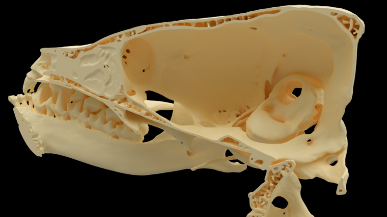
Figure 4 provides photorealistic rendered images of the bat skull, both intact as imaged and after digitally slicing away half of the volume to allow us to peek inside the sample non-destructively. These rendered images arise from STL models suitable for 3D printing or for use in biomimicry studies.
Conclusion
Micro-CT is a versatile technique that can image almost any sample type that both fits within the instrument and allows for X-ray energy to pass through without full attenuation. Within our current portfolio of instruments, the SkyScan 1273 proves to be a reliable workhorse for many laboratories around the country for ex-vivo imaging of animal and insect samples.
With essentially no sample preparation, the SkyScan 1273 allowed us to obtain a high-resolution dataset capturing the fine structural details within the bat skull in less than two hours of imaging time.
We hope you find this Image of the Month article informative and encourage you to subscribe to our newsletter and social media channels in preparation for the continuation of our Image of the Month series next month.
Scan Specifications
| Sample | Bat Skull |
| Detector | Flat-panel |
| Voltage (kV) | 80 |
| Current (µA) | 187 |
| Filter | 0.5 mm Aluminum |
| Voxel Size (µm) | 15 |
| Rotation Step (deg) | 0.2 |
| Exposure Time (ms) | 477 |
| Rotation Extent (deg.) | 360 |
| Scan Time (HH:MM:SS) | 01:56:23 |
These scans were completed on our SkyScan 1273 instrument at the Micro Photonics Imaging Laboratory in Allentown, PA. Reconstructions were completed using NRecon 2.0 while visualization and volumetric inspection of the 2D and 3D results were completed using DataViewer and CTVox. The dataset was converted to a volumetric model using Synopsys’ Simpleware software with the CAD add-on module (Synopsys, Inc., Mountain View, USA) before 3D rendering using Maverick Render Indie (Random Control, Madrid, Spain).
1. American Society of Mammalogists. (2026). Mammal Diversity Database, Version 2.5. https://doi.org/10.5281/zenodo.17033774