
Two Detectors, Different Views
Choosing between flat-panel and sCMOS imaging on the X4 POSEIDON Multi-Vision
Contents
- What is Multi-Vision?
- Why use two different detectors?
- What are the characteristics of the flat-panel detector?
- When is the sCMOS detector the better choice?
- Does the detector with more pixels always produce the better scan?
- How does sample size affect detector selection?
- Can both detectors be used within the same research project?
- What types of laboratories benefit from Multi-Vision?
- Can Multi-Vision be added later?
- How should a researcher choose between the two detectors?
- Matching the Detector to the Research Question
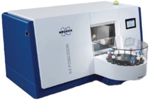
No single detector is optimal across the full range of sample sizes, fields of view, scan-time requirements, contrast requirements, and spatial resolution. Multi-Vision addresses that tradeoff by combining the flat-panel and sCMOS detectors within the transmission-source, Advanced Stage, and Best-Scan-Geometry architecture.
What is Multi-Vision?
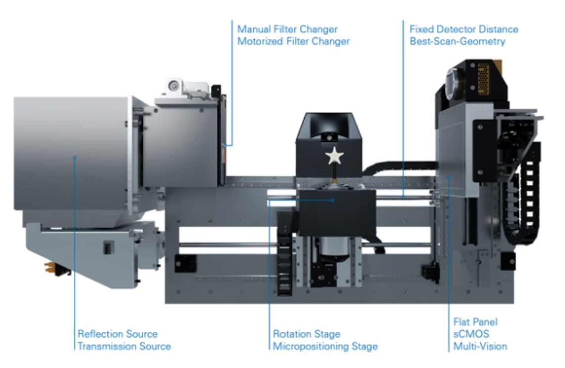
Multi-Vision is the dual-detector configuration of the X4 POSEIDON. It combines the transmission X-ray source, Advanced Stage, and Best-Scan-Geometry with both the sCMOS and flat-panel detectors, installed on the dedicated dual-detector mount with associated wiring.
Although an X4 can be configured with either detector individually, Multi-Vision requires this complete architecture. It allows researchers to select the detector and imaging geometry that best match the specimen and research question, while making the full performance range of both detectors practical within one system.
Why use two different detectors?
A detector that works well for one sample may not be the best choice for another. Larger specimens may require a larger field of view, while smaller samples or features may benefit from the sCMOS detector’s larger array of smaller pixels.
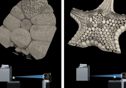
The two detectors allow the imaging approach to change with the sample. The Advanced Stage sets the sample position, which determines geometric magnification and field of view. Best-Scan-Geometry adjusts detector position; moving the detector closer to the X-ray source increases photon collection at the detector. That higher signal can be used either to reduce integration time and shorten the scan, or to maintain longer integration times to improve contrast differentiation and reduce noise.
What are the characteristics of the flat-panel detector?
The X4 flat-panel detector has 7 megapixels, with a 2800 × 2400-pixel array. Within Multi-Vision, it is particularly useful when a larger field of view, larger sample size, shorter scan time, or higher throughput is the priority.
It may be useful when the research question requires:
- A larger field of view or larger samples
- Shorter scan times
- Higher-throughput screening or comparison of multiple samples
- Improved contrast differentiation and reduced noise, especially when materials or tissues have similar X-ray attenuation
- Capturing time-dependent changes during in-situ studies
The key distinction in the X4 Multi-Vision configuration is that each detector has a different practical resolution limit. With the transmission source, the flat-panel detector is capable of approximately 3 µm resolution, with further improvement limited primarily by the detector architecture. The sCMOS detector, by contrast, can achieve approximately 2 µm resolution, at which point the 2 µm transmission-source focal spot becomes the limiting factor. The flat panel can therefore be the more practical detector when a larger field of view, faster acquisition, or higher signal is more important than resolving structures below approximately 3 µm, while the sCMOS detector is used when the highest available spatial resolution is required.
When is the sCMOS detector the better choice?
The scientific-grade sCMOS detector has 16 megapixels, with a 4096 × 4096-pixel array, and its individual pixels are smaller than those of the flat-panel detector. For a given sample size and imaging geometry, the combination of more pixels and smaller pixels supports higher spatial resolution when the rest of the system provides the required magnification and source resolution.
The detector does not create high-resolution system performance by itself. The transmission source, Advanced Stage, and Best-Scan-Geometry provide the source spot size, precision positioning, and imaging geometry needed to take advantage of the sCMOS detector’s larger array and smaller pixels.
It may be preferred when the study involves:
- Fine pores, cracks, fibers, or internal interfaces
- Small biological structures
- Thin coatings or walls
- Detailed morphology
- Closely spaced features that must be separated during analysis
Does the detector with more pixels always produce the better scan?
Not necessarily. A larger detector array and smaller detector pixels can support greater image detail, but neither determines image quality or system resolution by itself.
The useful resolution of a micro-CT scan also depends on the X-ray source spot size, geometric magnification, sample dimensions, material density, contrast, exposure settings, and reconstruction parameters. The smallest possible voxel size is not automatically the most appropriate choice for every sample.
Pixel count and pixel size are therefore inputs, not a ranking of detector quality. The appropriate choice is the detector and imaging geometry that provide the information required for the particular scan without unnecessary acquisition time or data volume.
How does sample size affect detector selection?
Sample size influences how much of the specimen must fit within the image and how much geometric magnification can be used. The Advanced Stage changes sample position relative to the X-ray source, which determines magnification and field of view. Best-Scan-Geometry then adjusts detector position to increase photon collection for the selected geometry.
The flat-panel detector is useful when a larger field of view or larger sample must be accommodated in a single scan. The sCMOS detector may be selected when the sample—or the feature of interest—is small enough to take advantage of its larger array and smaller pixels.
Detector selection should therefore account for both the overall sample dimensions and the size of the smallest feature that must be measured.
Can both detectors be used within the same research project?
Yes. Different stages of a project may have different imaging requirements.
A researcher might first use the flat-panel detector to examine the entire specimen, locate internal features, or screen a group of samples. The sCMOS detector could then be used when more detailed imaging is needed for selected specimens or structures.
This approach allows the imaging strategy to become more targeted as the study progresses.
What types of laboratories benefit from Multi-Vision?
Dual-detector capability can be valuable in laboratories that work with varied samples or support researchers with different imaging needs, including:
- Shared micro-CT and imaging facilities
- Multidisciplinary research laboratories
- Service laboratories analyzing samples for multiple clients
- Materials research and product-development groups
- Industrial laboratories combining screening with detailed failure analysis
- Life-science laboratories imaging specimens across different size scales
It can also be useful when a study includes both higher-throughput screening and detailed follow-up imaging.
Can Multi-Vision be added later?
Yes, but the practicality of a future Multi-Vision upgrade depends on the original X4 configuration.
Systems already equipped with the transmission source, Advanced Stage, and Best-Scan-Geometry offer the most direct upgrade paths. A High-Res configuration already includes the sCMOS architecture, so the flat-panel detector and required mounting hardware can be added. A transmission-source system configured with the Advanced Stage, Best-Scan-Geometry, and flat-panel detector can likewise be expanded by adding the sCMOS detector and the required mounting and support hardware.
Converting a standard reflection-source flat-panel system to Multi-Vision is a substantially larger and more expensive project. In addition to the sCMOS detector, the source, stage, detector-support hardware and bracket, wiring, and other components must be changed. Although the conversion is technically possible, it generally makes more financial sense later in the system’s service life, when replacing major components as part of the upgrade is easier to justify economically.
How should a researcher choose between the two detectors?
Detector selection should begin with the scientific question rather than the highest available specification. The main considerations include:
- Overall specimen dimensions
- Size of the smallest feature that must be resolved
- Required field of view
- Material density and X-ray attenuation
- Throughput and acquisition-time requirements
- Whether the study requires whole-sample screening, detailed structural analysis, or both
For a Multi-Vision user, the question is not simply, “Which detector is better?” It is, “Which detector and imaging geometry provide the information required for this particular scan?”
Matching the Detector to the Research Question
There is no single detector configuration that is optimal for every micro-CT scan. Imaging requirements change with the specimen, the feature of interest, and the purpose of the study.
By integrating the transmission-source architecture, Advanced Stage, and Best-Scan-Geometry with both flat-panel and sCMOS detection, Multi-Vision lets researchers trade among field of view, acquisition speed, contrast, and spatial resolution without relying on separate systems. Its value lies in selecting the detector and geometry that best answer the research question for each scan.
