
Jacob Waitkus
PhD Candidate, Chemical and Environmental Engineering
University of California, Riverside
Nanobiosensing, Nanomanufacturing, and Nanomaterials Laboratory (3N Lab)
Principal Investigator: Ke Du, PhD
Chemical and Environmental Engineering
Marlan and Rosemary Bourns College of Engineering
University of California, Riverside
Video YouTube link
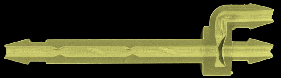
Researchers at the University of California, Riverside’s 3N Lab are developing a 3D-printed microfluidic device called the Ultra Rapid Anticlog Mixer (UltRA Mixer) for rapid mixing of solutions containing micro- or nanoparticles while limiting particle accumulation within the channel (Figure 1).
Device performance depends on enclosed internal features on the order of tens of micrometers. Micro-CT provided a nondestructive method for characterizing these features and assessing the fidelity of the as-printed geometry relative to the intended design.
Controlling Flow with Micron-Scale Baffle Geometry
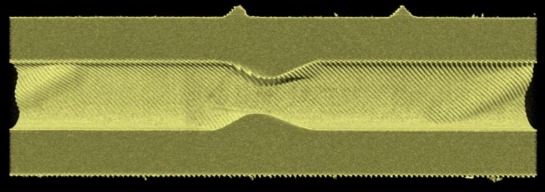
The UltRA Mixer is a passive microfluidic system in which mixing is generated by the internal channel geometry rather than by moving components. The device is fabricated from HTL resin using Projection Micro Stereolithography (PµSL) and incorporates a series of opposing baffle plate pairs within the mixing channel. The baffle geometry redirects the flow toward the center of the channel to promote mixing.
This configuration was developed in part to address particle accumulation in microfluidic systems. Nanoparticles and microparticles can collect around corners and other internal features, altering local flow conditions, reducing mixing reproducibility, and potentially obstructing the channel. The UltRA design was intended to maintain rapid mixing while minimizing regions where suspended particles could accumulate.
Reducing Mechanical Stress During Protein Capture
The researchers evaluated the UltRA Mixer for an antibiotic-resistance application involving penicillin-binding proteins PBP1a and PBP3. These proteins interact with β-lactam antibiotics including penicillin and cephalexin. In the experimental system, the proteins were captured on micron-scale magnetic beads to study their interactions with the antibiotics.
Initial experiments used manual pipette mixing, but repeated pipetting resulted in inconsistent protein capture. Comparative experiments indicated that pipette mixing disrupted protein structure and reduced target-antibiotic binding and capture efficiency.
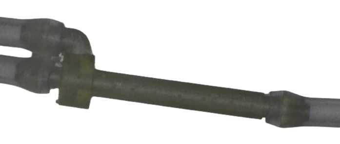
The UltRA Mixer provided an alternative method for maintaining contact between the magnetic beads and proteins while reducing the mechanical forces associated with manual pipetting. Surgical tubing linked the mixer’s inlet and outlet ports to peristaltic pumps, allowing flow rates and ratios to be controlled while maintaining effective mixing and protein capture (Figure 3).
Verifying Micron-Scale Fabrication
Performance of the mixer depends on accurate reproduction of the CAD-designed baffle geometry within the 3D-printed channel.
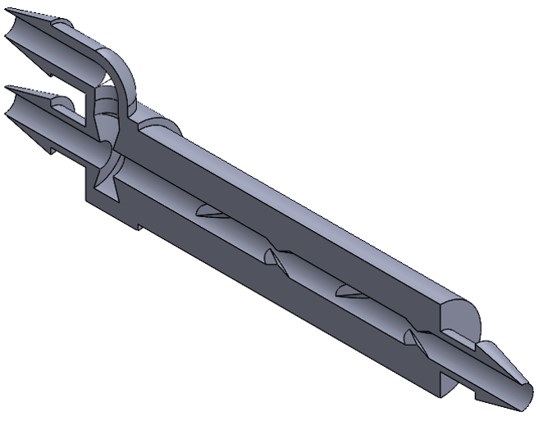
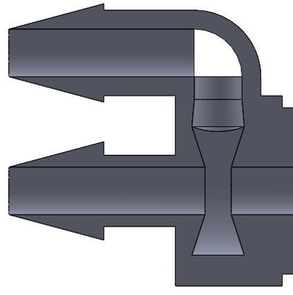
The UltRA Mixer was designed by Jacob Waitkus in SolidWorks (Figures 4, 5) and fabricated using a BMF P140 printer. The 20 µm specification of the printing process represents both the minimum feature resolution and the smallest achievable layer thickness. At this scale, fabrication variations can influence fluid behavior. Incomplete baffle formation, layer misalignment, residual resin, air pockets or partial obstruction of the channel could modify the internal flow field.
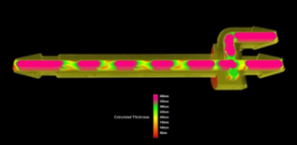
Because these structures are enclosed within the finished device, direct inspection would require destructive sectioning. Micro-CT provided a nondestructive method for characterizing the internal geometry of the completed device. The scans were used to evaluate baffle formation and spacing, inspect the chamber where the two inlet streams converge, identify residual resin or other fabrication defects, and characterize surface features associated with the individual print layers.
High-magnification scans of individual baffle pairs were used to assess print-layer uniformity and identify potential misalignment, defects and air pockets. Imaging at approximately 800 nm resolution was also used to characterize surface roughness within the mixing channel. A second series of scans at approximately 3 µm resolution visualized the internal structures throughout the fluid pathway, from the inlets through the mixing channel to the outlet. Micro-CT thereby provided structural characterization of the as-printed internal geometry and data relevant to subsequent experimental and computational analyses.
Incorporating As-Printed Geometry into Simulation
Micro-CT data also allowed the researchers to compare the fabricated mixer with the original CAD geometry and support simulations of protein mixing in highly viscous environments.
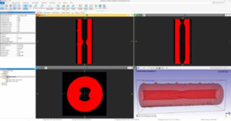
By incorporating observed characteristics of the as-printed structure—including surface features and grooves between print layers—the computational models could more closely represent the devices used experimentally. This provided a framework for relating the designed geometry, as-printed structure, and experimentally measured fluid behavior.
Evaluating Clogging and Protein Capture
Experimental testing indicated that baffle configuration influenced particle accumulation. UltRA Mixer channels were compared with configurations containing fewer baffle plate pairs, and microparticle behavior within the channels was observed. The UltRA configuration demonstrated reduced clogging during the assay.
Comparisons of pipette and UltRA mixing also demonstrated improved target capture with the UltRA approach. A fluorescent β-lactam analog was used to evaluate capture on the protein-functionalized microparticles by measuring the concentration remaining in solution following mixing and incubation.
Together, these experiments evaluated two aspects of device performance: the ability to mix particle-containing solutions while limiting particle accumulation, and the ability to support protein capture while reducing the mechanical stresses associated with manual pipetting.
Application to Biomarker Capture and Sensing
The UltRA Mixer is part of a broader research program within the 3N Lab focused on biomarker capture and sensing using micro- and nanoscale devices. The interdisciplinary group combines nanomanufacturing, nanostructured materials, and automated microfluidic systems to improve the sensitivity and specificity of biosensing platforms.
Current projects include methods for detecting antibiotics and antibiotic-resistance biomarkers at trace concentrations, with the goal of developing approaches that can support more rapid identification of clinically relevant biomarkers and help inform treatment selection for drug-resistant infections.
For the UltRA Mixer, micro-CT enabled characterization of the as-printed internal geometry, providing a structural basis for interpreting experimental performance and informing computational models.
Research Credits
University of California, Riverside:
Jacob Waitkus — PhD Candidate, Chemical and Environmental Engineering
Ke Du, PhD — Principal Investigator, 3N Lab
Yaneth Larios — Undergraduate Researcher, Bioengineering
Negin Bahadori — PhD Candidate, Chemical Engineering
Henry Yuqing — PhD Candidate, Mechanical Engineering
Lawrence Livermore National Laboratory:
Sankar Narayanasamy, PhD — Staff Scientist and Principal Investigator
Micro-CT scans were completed on Bruker’s X4 POSEIDON™ modular micro-CT system at the Micro Photonics Imaging Laboratory in Allentown, PA.
Scan Specifications
| Scan | High Res FOV | Overview |
| Detector | sCMOS | sCMOS |
| Voltage (kV) | 50 | 50 |
| Current (µA) | 40 | 100 |
| Filter | None | None |
| Voxel Size (µm) | 0.8 | 3 |
| Rotation Step (deg) | 0.2 | 0.3 |
| Exposure Time (ms) | 1466 | 595 |
| Rotation Extent (deg.) | 360 | 360 |
| Scan Time (HH:MM:SS) | 03:17:08 | 01:21:19 |