

With more than 35,000 species of ray-finned fishes and about 500 sharks identified worldwide, fish demonstrate more species diversity than all other kinds of vertebrates. A unique project using micro-CT scanning is changing the way scientists, teachers, students, and amateur ichthyologists will be able to study any fish from around the globe. This innovative use of micro-CT technology and scientific collaboration will enable anyone to look at the fine details of any fish online or produce an exact replica using 3D printing.

University of Washington professor of biology and aquatic and fishery sciences, Adam Summers has undertaken the daunting task of scanning and digitizing each species of fish through collaboration with other scientists, creating high-resolution, 3D images that will be available for free online. “These scans are transforming the way we think about 3D data and accessibility,” said Summers. “It’s been so fun to throw this data up on the web and have people actually use it.”
Adam Summers runs the Comparative Vertebrate Biomechanics Lab, which houses a SkyScan 1273 micro-CT scanner at Friday Harbor Laboratories, a research and educational facility.
We talked with Graham Short and Meghana Binraj about the micro-CT scans they have done in Adam Summers’ lab, and they have shared some stunning images of their work.
Can you expand on what you each are focused on in your research?
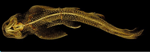
Graham Short’s research is centered on the taxonomy and systematics of fishes in the family Syngnathidae, which includes seahorses, pipefish, and seadragons. His current focus is on describing new seahorse species across the Indo-Pacific region.


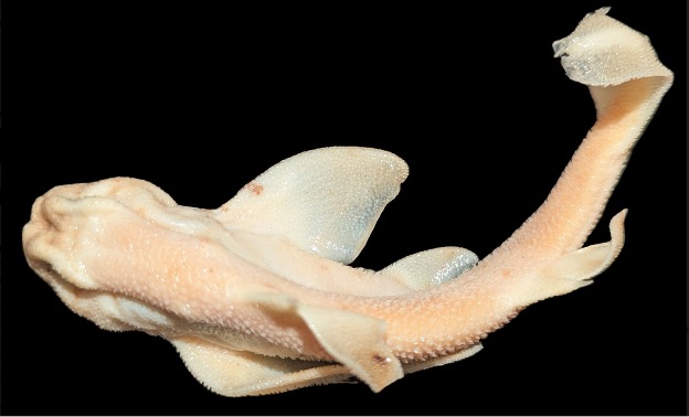
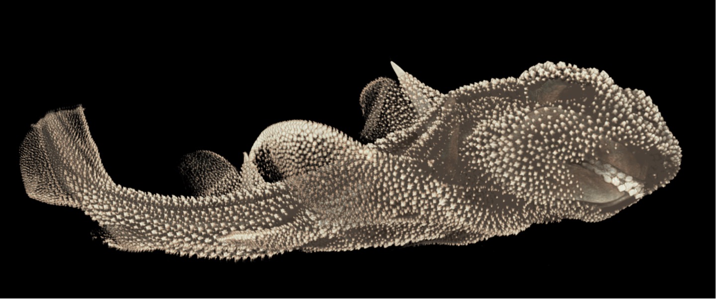
Meghana Binraj specializes in sharks and X-ray taxonomy and conservation in India. She was a 2024 Fish Class student in 2024 at Friday Harbor Laboratories where she was introduced to the science of micro-computed tomography (micro-CT) scanning technology that allows for detailed, high-resolution visualization of internal structures like bones (cartilage in case of sharks and rays), organs, and tissues within a fish, enabling early career researchers like her to measure and analyze their size, shape and spatial relationships in three dimensions.
What is important about this research today, and what are you hoping to achieve through your work?
Graham Short: Globally, there are 46 recognized species of seahorses, but many of these species actually represent distinct, cryptic species complexes. Accurately assessing the diversity of seahorses will lead to better-informed conservation and management strategies.
Meghana Binraj: In India, the diversity of sharks is not well understood. Conducting thorough diversity assessments will enhance shark conservation efforts in the region. The micro-CT scanning technique is crucial in contemporary research as it enables detailed visualization of the morphology of sharks and rays without invasive procedures. In India, identifying sharks from fishery landings can be challenging since specimens often lack critical external features. Many species also have internal characteristics that could be key to accurate identification, especially for those that are under-researched and may even be new to science.
By conducting comprehensive diversity assessments using micro-CT, we can explore these hidden morphological traits and answer important taxonomic questions. This advanced imaging tool allows me to visualize species diversity in a non-invasive way, ultimately enhancing conservation efforts in the region. My goal as an early-career researcher is to take these skills back home, apply them to our local fisheries, and contribute to effective conservation strategies tailored to the unique needs of India’s shark and ray populations.
How are you using micro-CT technology in your research?
Graham Short: It was thanks to Adam, who told me about the necessity to use the micro-CT scanner to examine fish in detail. It was a game changer for me since I was relying solely on microscopy. When I learned how to use the scanner, I was amazed at the incredible detail of the skeletal features of fish.
I live in Australia half the year. Otherwise, I’m in Friday Harbor during the summer months. I introduced myself to Adam ten years ago since I’ve always been curious about the Friday Harbor Marine Labs. That first time we met he told me about the micro-CT scanner, it’s totally changed my methods for taxonomic research.
Micro-CT scan technology allows for the detailed examination of subtle morphological features in seahorses that are often overlooked by conventional methods like microscopy and X-rays. Scanning the entire specimen enables the generation of high-resolution images of specific body parts, such as the head, which helps distinguish new species from known ones.
Meghana Binraj: As a newcomer to the world of micro-CT technology, I’m currently focused on acquiring skills to visualize and image sharks, rays, skates, and chimaeras. I feel incredibly fortunate to have the opportunity to learn with the SkyScan 1273, and I want to thank Graham for allowing me to shadow him as I pick up skills and conduct my own scans under his supervision. I’m also grateful to Adam for the wonderful opportunity to spend time in his lab, which has been invaluable for my learning experience.
I’ve completed a few quick scans lasting around 10 minutes, as well as longer scans that take four to six hours, and both have yielded impressive results. I’m particularly excited about the upcoming Dice CT scans, which will allow me to visualize soft tissues. Every time I process the CT scans and observe the high-resolution images, I’m in awe of the intricate details that emerge. This new skill is enabling me, as an early-career shark researcher, to approach these species from an entirely new perspective and ask questions I wouldn’t have considered otherwise.
This experience has highlighted for me how each new skill in science can play a pivotal role in shaping our understanding and sparking innovative inquiries. I look forward to the insights that will arise as I continue to develop my expertise in this exciting field.
For sharks, CT images reveal features like teeth structures that are critical in differentiating closely related species.
What has been the most important discovery or unexpected outcome in your research?
Graham has described several new species of seahorses and pipefish using micro-CT scan data. Inspired by Adam Summers, who emphasized the importance of micro-CT scanning for detailed fish examination, Graham’s use of this technology has led to the discovery of novel morphological features in seahorses. This approach has set a new standard in seahorse taxonomy.
Are there any relevant publications we could include links for?
Graham described a new species from South Africa, with the publication featuring CT scan images of the species. You can access the paper here: A New Record and Species of Pygmy Pipehorse of the Genus Idiotropiscis from South Africa.
Credits:
Graham Short (seahorse images)
Research Associate, Burke Museum, University of Washington
Ichthyology Research Fellow, Australian Museum
Meghana Binraj (shark images)
Species and Spaces Foundation, India
Minorities in Shark Sciences, USA
With special thanks to Adam Summers for his inspiration.
Comparative Vertebrate Biomechanics Lab, Friday Harbor Laboratories
School of Aquatic and Fisheries Sciences
University of Washington
620 University Drive
Friday Harbor, WA 98250
If you are interested in being featured in a future Researcher Spotlight, please contact ann@microphotonics.com. We love to hear how our instruments are being used in the field!
