Les Watling, PhD
Professor Emeritus, School of Life Sciences, University of Hawaii at Mānoa, and Emeritus Professor of Oceanography, University of Maine’s Darling Marine Center
Megan L. Porter, PhD
Professor and Associate Director of Research, School of Life Sciences, University of Hawaii Dr. Porter is responsible to the micro-CT scanning.
To highlight various applications of micro-CT technology, Micro Photonics is featuring investigators working in both life science and materials science. We continue this series with an interview with Les Watling, who uses micro-CT technology in his biosciences research.
I know you study the taxonomy, biogeography, ecology, and reproduction of deep-sea octocorals. Can you expand briefly what you are focused on in your research?
In this project I am trying to understand the mechanical design of the octocoral polyp. In many respects the polyps are considered to be quite simple, being made of only two cell layers and not having any complex organs of any kind. But within those limits there are many different forms, all seemingly focused around providing defense for the polyp. That is, some polyps do not contract much when bothered and just pull the tentacles over the mouth area. The top of the polyp is then protected by the projecting needle-like sclerites, as in the images shown here. But at the other extreme are polyps that either contract strongly or tuck the tentacles into a funnel-like feature at the top of the polyp. The one internal structure that all octocorals have, the pharynx, then needs to collapse, usually in a form like a contracting bellows.
For the most part my samples have come from seamounts in the North Atlantic Ocean but my research is expanding to cover seamounts and island slopes in the Hawaiian and nearby areas. My colleagues and I are particularly interested in the modes of dispersal of these animals, their long-term evolutionary history, and their relationships to deep ocean water masses. Deep-sea octocorals are also home to a variety of smaller invertebrates. I am interested in how these relationships develop and are maintained. Because these animals live at depths that are accessible only by submersible or remotely operated vehicles, much of what is known has to be gained by inference. We examine closely the bodies of the commensals for clues to their roles in these relationships.
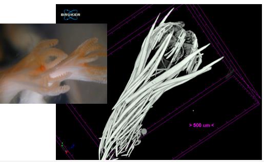
The live image of the octocoral polyp is quite ethereal and beautiful. Are they hard or soft?
I completely agree, sometimes the octocoral polyps are really spectacular, but I do like your use of the word ethereal (Figure 1). The tissue of the polyp is soft and thin, but the calcitic structures, that sometimes look like they are made of glass, are hard — well, as hard as calcite can be. And they are brittle and easily broken when handling the specimen.
How are you using micro-CT technology in your research?
To understand how the polyp is designed and functions, one option is to do careful dissections of the polyp body, then fix and dry the dissected specimen and look at it with a scanning electron microscope (SEM). But with micro-CT it is possible to see the internal structures without dissection. To image the soft and delicate tissue on the inside of the polyp, we soaked the specimen in a solution of osmium tetroxide and then embedded the polyp in epon, a form of plastic. That is a standard method of preparation of the polyp for transmission electron microscopy (TEM), but we discovered it to be ideal for micro-CT work.
Micro-CT Octocorals Image Gallery
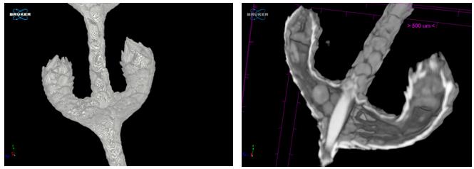
Figure 2 shows two polyps of the octocoral species Thouarella grasshoffi from Manning Seamount, NW Atlantic, collected at 1461 m depth, showing internal anatomy. These polyps are very small so some of the internal features of larger polyps are missing and the number of eggs is small (one egg can be seen near the base of the polyp on the left).
The image at the right (Figure 2) is the same specimen, but illustrating the armoring of the polyp by calcareous plates secreted by the epidermal cells. The lines appear as contours on the plates (and to be honest, I can’t remember how I generated them, but it was some combination of Luminance and Opacity).
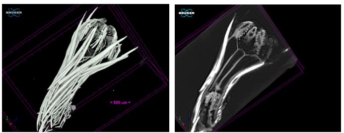
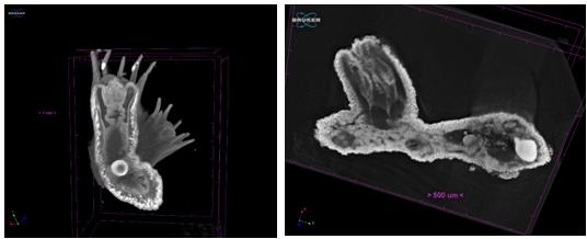
Changing the opacity allows us to see the arrangement of the calcareous sclerites, which are used to support the tissue of the polyp shown in side view (Figure 3, left). The size, shape, and arrangement of the sclerites are important features used in the taxonomic description of octocorals.
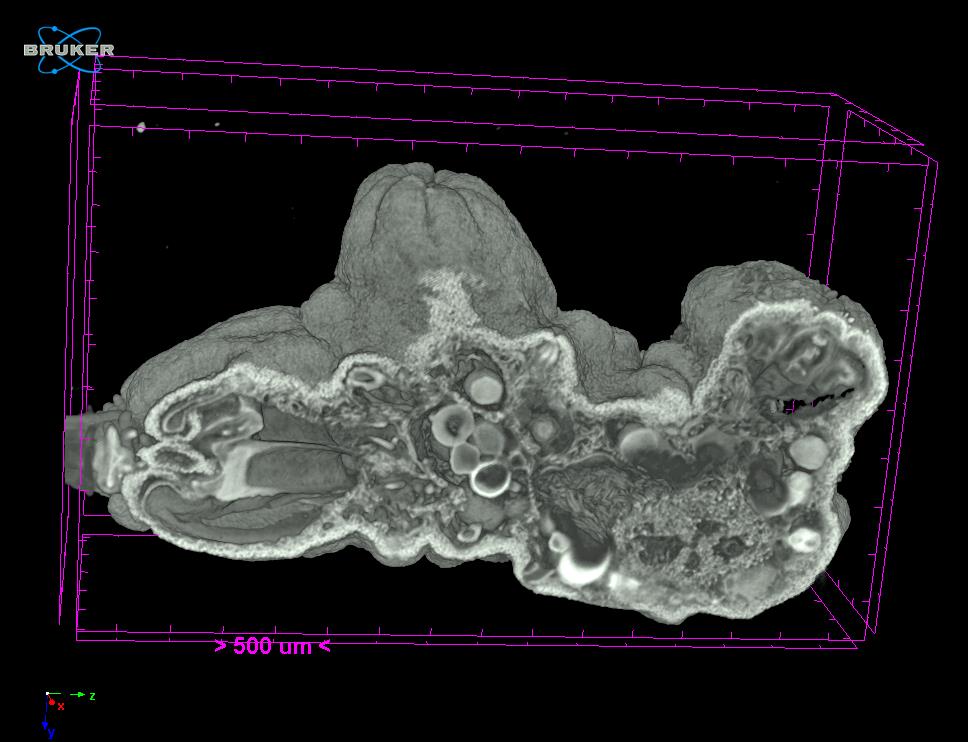
The long cut with pharynx (Figure 3, right) shows a single polyp of a new, yet to be described species of octocoral collected from Kelvin Seamount, NW Atlantic, at 2015 m depth, showing the internal structure, especially the tentacles at the top of the polyp, and the mesenteries near the base. The white objects on the side are calcareous sclerites.
What is important about this research today, and what are you hoping to achieve through your work?
We have been conducting a survey of octocoral polyps from different taxonomic families to see what the diversity of morphology is like. I think the most interesting discovery so far is that there is a very wide range of morphologies, especially considering that this group of animals has a so-called simple body plan. From a functional perspective, the polyp needs to be able to gather food, grow, reproduce, and contract in some way to defend itself. All this while sitting immobile on the skeletal axis of the colony. To feed it extends the polyp body and tentacles into the water where particles can be intercepted and moved to the mouth. To reproduce it needs to be able to make either eggs or sperm, and it needs a way to either dump the eggs and sperm into the water where fertilization occurs, or perhaps the females somehow catch the sperm from the water. None of that is known. And then, when disturbed or when something is trying to feed on it, the polyp has various defensive measures such as contraction of part, like the tentacles, or all of the body. The design of the polyp has to balance all those needs, and we are looking to see how that is done. Without getting into a long zoology lesson, evolution in this group has come up with a diversity of solutions.
We have nearly finished our survey of the groups of octocorals where we have well-prepared specimens, and we are also working on the taxonomy of these species because they are all new to science and need describing and naming.
More Information:
To contact Les Watling, please email: watling@hawaii.edu.
If you are interested in being featured in a future Researcher Spotlight, please contact ann@microphotonics.com. We love to hear how our instruments are being used in the field!