Micro-CT to Visualize Implants and Surrounding Bone

Aatif Jabbar, BS;  Nahir Habet, MSc; Sarah Romereim, PhD; Bailey Fearing, PhD
Musculoskeletal Institute
Atrium Health
Charlotte, NC

Figure 1: Micro-CT reconstruction of a femoral implant in a rabbit knee model.


There are about one million total hip arthroplasties and total knee arthroplasties performed in the United States every year, a number that is expected to grow with an increasingly aging population. Prosthetic Joint Infection (PJI) is a complication following total arthroplasty in about 1-2% of these surgeries and is responsible for about 30-40% of all failures in revision. Research to improve treatment and prevention for PJI focuses on new antibacterial coatings, surface designs, or other compounds to protect implants, with or without antibiotics.

The Orthopaedic Engineering Research Laboratory at Atrium Health applies basic engineering principles, biomechanical evaluation techniques, and new biotechnologies to orthopaedic surgery to improve clinical outcomes. The lab combines experimental mechanical studies of implantable devices and the development of rapidly translatable and novel infection interventions to address implant-associated infections. High‑resolution, non-destructive micro-CT is a key tool in their orthopaedic research program.

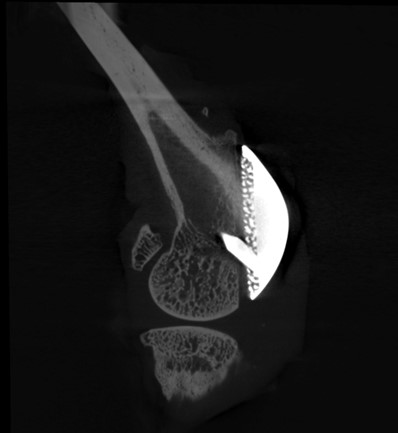
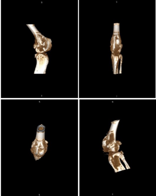
Micro-CT Imaging of Implant Osseointegration in Rabbit Models

High‑resolution micro-CT enables the non‑destructive, 3D visualization of implants and surrounding bone over time. The images presented here showcase the use of micro-CT to study periprosthetic joint implantation and osseointegration in rabbit models, allowing tracking of implant positioning, bone remodeling, and early indicators of bone integration at multiple time points.

Below are micro-CT reconstructions of a femoral implant in a rabbit knee model. High-resolution imaging enables tracking of implant position and progressive changes at the implant-bone interface over time.

Figure 2: Multi-planar reconstruction view of femoral implant in rabbit knee model visualizing placement of implant within femoral head
Figure 3: Multi-planar reconstruction view of femoral implant visualizing fit within
medullary cavity
Figure 4: Multi-planar reconstruction view of femoral implant and adjacent tibia

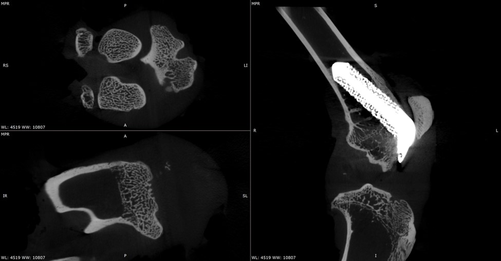
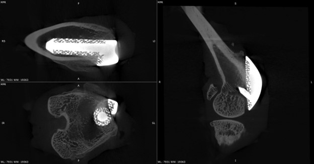
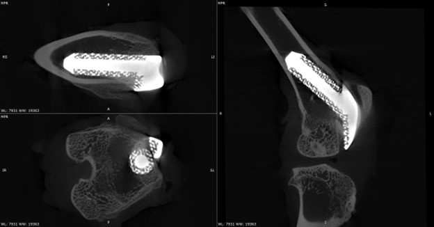
Temporal Assessment of Implant-Bone Interactions

Micro-CT imaging allows repeated scanning of the same implanted joint, providing a powerful means to observe temporal changes in bone architecture without disrupting the tissue. These scans allow monitoring of how the surrounding trabecular and cortical bone adapts to the presence of the implant, capturing subtle changes in bone volume, density, and structure as healing progresses. This approach enables precise comparison of early versus later stages of implant integration within the rabbit knee.

Figure 5: 3D reconstruction planar views of femoral implant and bone interface


Orthopaedic Research at Atrium Health

The study shown here demonstrates efforts to develop preclinical models that allow testing of implants with infection challenges and overall outcomes. But much of the work from Atrium also focuses on investigating implantable device selection and optimization, as well as surgical techniques by comparing mechanical testing of multiple devices that have been developed for similar clinical applications. These studies are often performed with various orthopaedic implants, including installation and fixation of fractures and joint replacement devices in primary or revision surgeries, and anchoring devices for soft tissue reconstructions involving tendons, ligaments or joint capsule segments. Data collected is often used to evaluate initial stabilization or long-term fatigue performance and studies may utilize either surgical patient samples, synthetic or cadaveric specimens, and/or animal models. These studies typically involve the evaluation of stresses, strains, load distributions, micro motion, pressure or bone ingrowth at the fracture site or device / bone interface. The laboratory houses an extensive array of electronic instrumentation and test systems for performing experimental biomechanics studies, and micro-CT is a key tool within this lab research program.

Additional research involving micro-CT and the intersection of surgical implants and bone morphology:

Micro-CT Imaging and Mechanical Properties of Ovine Ribs

Effects of local and systemic ketorolac application on rat femur fracture healing

Development of a small animal bone-anchored limb replacement model for infection interventions – PMC

All-suture anchor pullout results in decreased bone damage and depends on cortical thickness

Compressive Strength of Midfoot Fusion Nail vs Midfoot Fusion Bolt and Role of Subtalar Fusion in Midfoot Charcot Fixation Model

To contact the team, please email: Nahir.Habet@atriumhealth.org or Aatif.Jabbar@advocatehealth.org

Images scanned on a Bruker SkyScan 1273 micro-CT © 2026 Atrium Health.

If you are interested in being featured in a future Researcher Spotlight, please contact ann@microphotonics.com. We love to hear how our instruments are being used in the field!

Related Products

Related Articles

US Partner Form


    Coming Soon:

    Customer access to tips and instructional videos, method notes, tutorials, application notes, and other content to support your research.

    Employment

    Our people are the key to our success.

    We seek and employ talented scientists who have the curiosity and knowledge to offer intelligent collaboration with the researchers who are our customers. We don’t have any openings but would be happy to receive resumes to hold on file.


    Please submit resumes to: info@microphotonics.com

    Micro Photonics Inc.
    1550 Pond Road, STE 110
    Allentown, PA 18104

    Events

    CONFERENCES

    ORS 2026 Annual Meeting
    March 27 – 31, 2026
    Charlotte Convention Center
    Charlotte, NC
    2026 Annual Meeting – ORS

    Preclinical Imaging Consortium
    Philadelphia, PA
    April 13-15, 2026

    AALAS National Meeting
    Houston, TX

    Oct. 25-29, 2026
    AALAS

    Privacy Policy

    What information do we collect? We collect information from you when you register on our site, place an order, subscribe to our newsletter or fill out a form. When ordering or registering on our site, as appropriate, you may be asked to enter your: name, e-mail address, mailing address, phone number or credit card information. You may, however, visit our site anonymously.
    What do we use your information for? Any of the information we collect from you may be used in one of the following ways:

    ; To personalize your experience (your information helps us to better respond to your individual needs).

    ; To improve customer service (your information helps us to more effectively respond to your customer service requests and support needs).

    ; To process transactions. Your information, whether public or private, will not be sold, exchanged, transferred, or given to any other company for any reason whatsoever, without your consent, other than for the express purpose of delivering the purchased product or service requested.

    ; To send periodic emails .The email address you provide for order processing, will only be used to send you information and updates pertaining to your order.Note: If at any time you would like to unsubscribe from receiving future emails, we include detailed unsubscribe instructions at the bottom of each email.

    Terms and Conditions

    Terms of Service for www.microphotonics.com

    Introduction. Welcome to www.microphotonics.com. This website is owned and operated by Micro Photonics Inc. By visiting our website and accessing the information, resources, services, products, and tools we provide, you understand and agree to accept and adhere to the following terms and conditions as stated in this policy (hereafter referred to as ‘User Agreement’), along with the terms and conditions as stated in our Privacy Policy (please refer to the Privacy Policy section below for more information).

    This agreement is in effect as of October 25, 2018.

    We reserve the right to change this User Agreement from time to time without notice. You acknowledge and agree that it is your responsibility to review this User Agreement periodically to familiarize yourself with any modifications. Your continued use of this site after such modifications will constitute acknowledgment and agreement of the modified terms and conditions.

    Responsible Use and Conduct. By visiting our website and accessing the information, resources, services, products, and tools we provide for you, either directly or indirectly (hereafter referred to as ‘Resources’), you agree to use these Resources only for the purposes intended as permitted by (a) the terms of this User Agreement, (b) the terms of any applicable confidentiality, non-disclosure, or other agreement between you and a third party, and (c) applicable laws, regulations and generally accepted online practices or guidelines.

    Call Now Button