This Researcher Spotlight highlights the work of Joseph Wallace, PhD, who uses micro-CT and DEXA technology in his bone research. The lab incorporates co-morbidity models that reflect clinical patient complications, such as diabetes and chronic kidney disease (CKD) that impact bone health and increase bone fracture risk.
Joseph Wallace, PhD
Associate Vice President of Research Development
Professor of Biomedical Engineering
Weldon School of Biomedical Engineering
Purdue University
Dyann Segvich
Senior Laboratory Operations Specialist
Weldon School of Biomedical Engineering
Purdue University
Can you expand briefly on your focus in your research?
Providing the body’s framework, bones are essential for support and protection. They offer a store of essential minerals that can be utilized when needed, especially calcium and phosphorus. Bones also undergo a continuous remodeling throughout life that requires a delicate balance of resorption and formation to repair damage and keep them robust. Understanding bone turnover elucidates possible avenues for therapeutic interventions to prevent bone loss and fracture. In addition to turnover, investigating the major protein found in bone, collagen, allows for unique therapeutic avenues. The laboratory of Dr. Joseph Wallace at Purdue University focuses on understanding bone biology and mechanobiology from the molecular level all the way to the tissue level of organisms. To do this, we utilize a variety of tools including cellular and murine models.
What is important about this research today, and what are you hoping to achieve through your work?
With age and disease, bone quantity and tissue quality are compromised, which drastically increases fracture risk. In our research, we explore ways to enhance bone growth as well as improve the quality of pre-existing and newly formed bone. We focus on diabetes, chronic kidney disease (CKD), and other disorders that alter bone health. The rising percentage of people diagnosed with these diseases makes the importance of this work more critical now than ever. The goal of our team is to uncover how disease transforms bones in conjunction with looking for anabolic and pharmaceutical interventions that can improve bone health and fracture resistance and ultimately be translated to the clinic.
What makes your research unique?
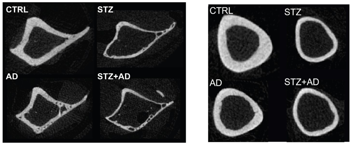
Often, research focuses on a single disease that impairs bone health. Our lab incorporates co-morbidity models that reflect clinical patient complications. For instance, diabetes and chronic kidney disease (CKD) are two ailments that impact bone health and increase bone fracture risk. In the US, close to 1 in 3 adults with diabetes also suffer from CKD. This is known as diabetic kidney disease (DKD). Our lab developed a novel murine model that found DKD bones to have greater detriments in both cortical and trabecular bone compared to either disease alone. This work was continued and showed that a combined drug treatment with raloxifene, a selective estrogen receptor modulator, and romosozumab, an anabolic, leads to the greatest improvements in bone mass and tissue quality in the DKD model.
How are you using micro-CT technology in your research?
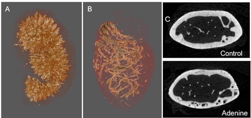
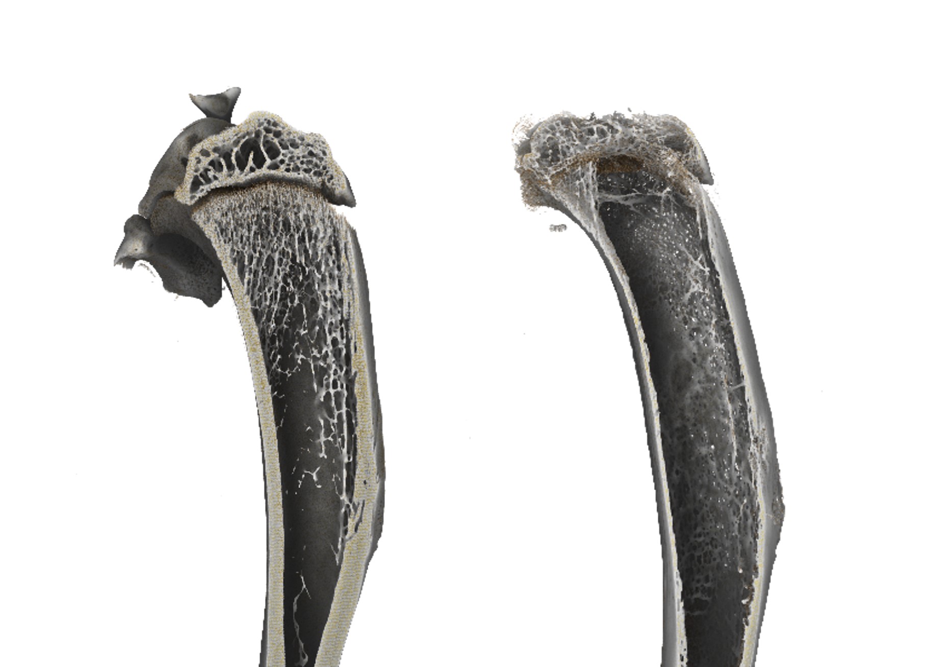
The Bruker Skyscan 1272 is the cornerstone of our research. It enables us to generate 3D renderings of bone and soft tissue, giving a glimpse of the geometry and morphometry of tissue samples. We focus on analyzing both the cortical and trabecular bone and look for phenotypic differences in our experimental groups. Scanning bones ex-vivo also gives us the ability to determine tissue mineral density and porosity of our samples. Since this technique is non-destructive, we can use the same samples for other chemical and mechanical tests following micro-CT imaging. Soft tissue, like kidney and heart, are also scanned to look for damage or vascular calcification.
How are you using DEXA technology in your research?
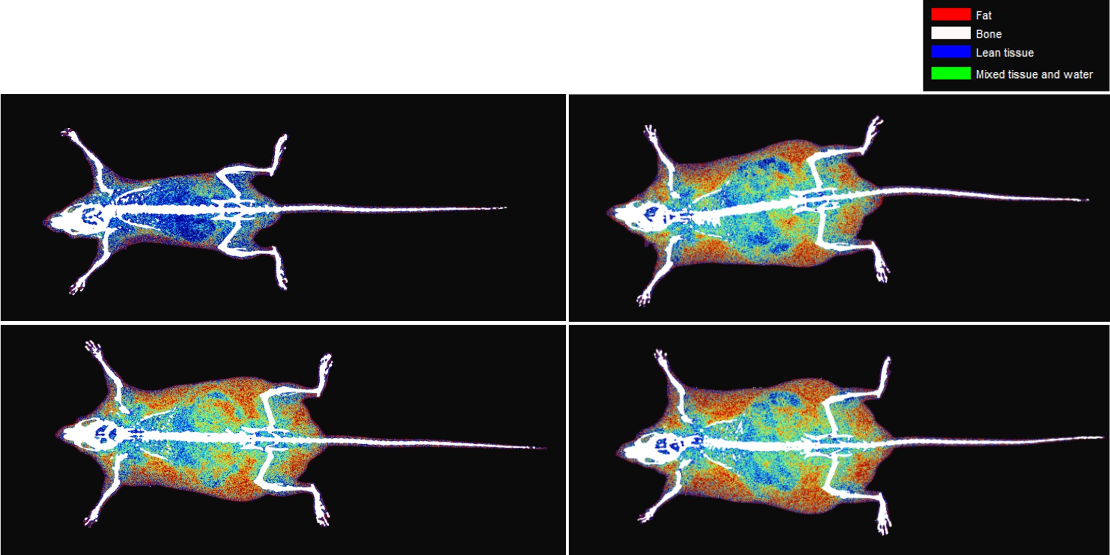
The InAlyzer2 DEXA (Medikors) allows us to collect longitudinal data from live animals that elucidates the changes happening throughout a study on the same cohort of mice. For our diabetic models, we can track the body composition over time after the introduction of a high fat diet. In addition to ex-vivo BMD bone measurements with the micro-CT, tracking changes to bone mineral density with in-vivo DEXA scanning also gives insight into disease progression and severity. This type of imaging also provides a clinical surrogate since this is the same type of imaging performed in human patients when diagnosing bone disease or assessing treatment efficacy.
Are there any images that you could share illustrating results using micro-CT and DEXA with your applications?
Selected Publications:
Damrath, John G et al. “A novel murine model of combined insulin-dependent diabetes and chronic kidney disease has greater skeletal detriments than either disease individually.” Bone vol. 165 (2022): 116559. doi:10.1016/j.bone.2022.116559
Kohler, Rachel et al. “Combined Romosozumab and Raloxifene treatment targets impaired bone quality in a male murine model of diabetic kidney disease.” Bone vol. 194 (2025): 117415. doi:10.1016/j.bone.2025.117415
Reul, Olivia N et al. “Practical Considerations for the Design, Execution, and Interpretation of Studies Involving Whole-Bone Bending Tests of Rodent Bones.” Journal of visualized experiments : JoVE ,199 10.3791/65616. 1 Sep. 2023, doi:10.3791/65616
Website: https://engineering.purdue.edu/BBML/index.html
All photos: © 2025 Joseph Wallace, PhD, Weldon School of Biomedical Engineering, Purdue University. All rights reserved.
__________
If you are interested in being featured in a future Researcher Spotlight, please contact ann@microphotonics.com. We love to hear how our instruments are being used in the field!