
The propagation of plants through their clever use of seeds is well studied in academia and useful in biomimicry applications. While most seeds have evolved distinct characteristics to help ensure the survival of each species, pods left behind that are full of seeds after the blooms fall from a snapdragon plant have a unique, almost face-like, appearance. In this article we present both a high-resolution dataset of the entire seed pod along with an even higher resolution dataset of an isolated individual seed, both acquired on the SkyScan 1272 high-resolution desktop micro-CT. The ability to image a sample at multiple resolutions helps researchers balance the fidelity of their data with the necessary acquisition time to capture the results.
X-Ray Microscopic Imaging of Plant Materials
We examined an entire dry seed pod from a snapdragon bloom at an isotropic voxel size of 8µm and then acquired a second dataset of an isolated seed near the limit of resolution of the SkyScan 1272 at an isotropic voxel size of 900nm.
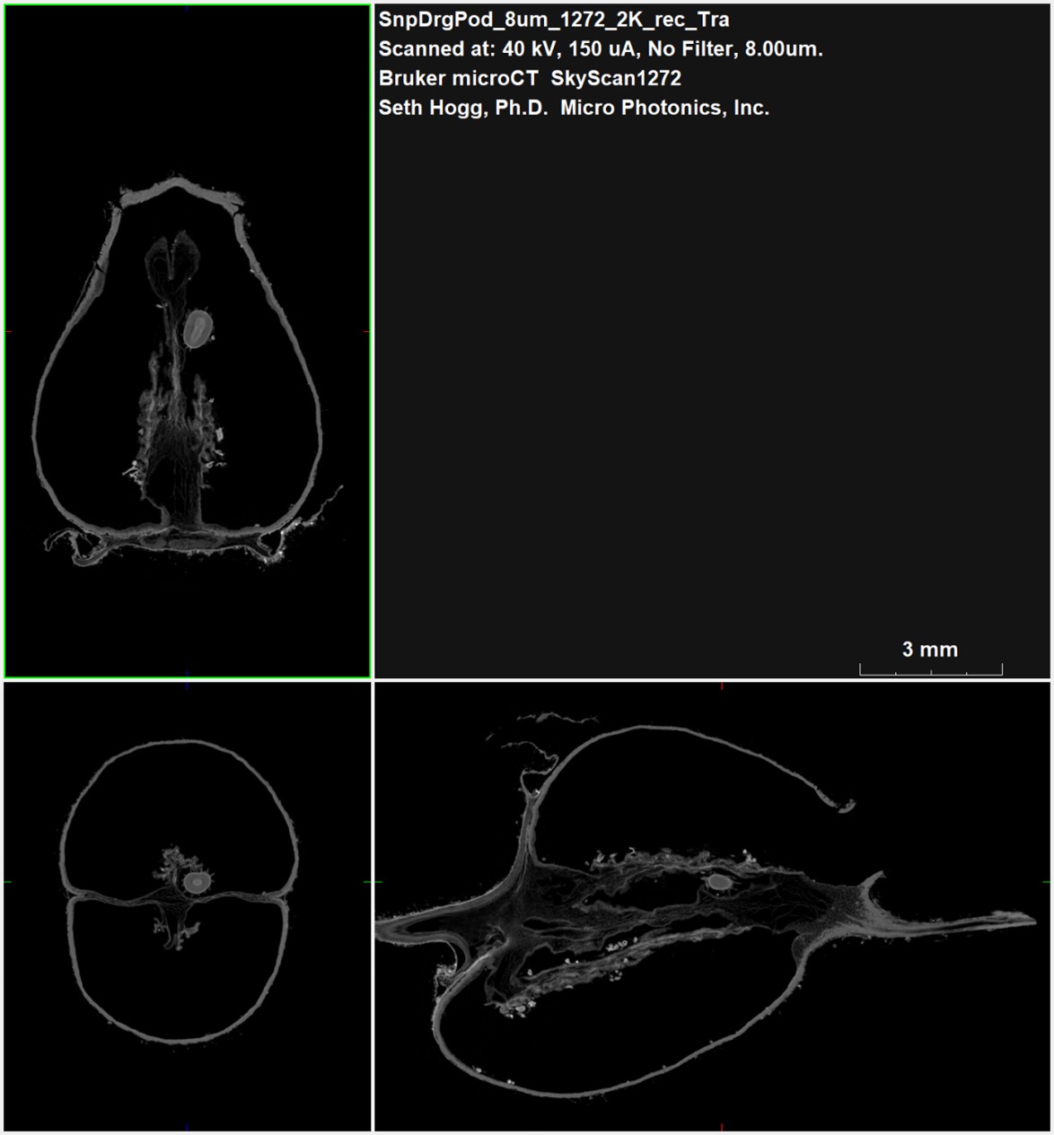
As shown from Bruker DataViewer in Figure 2, the seed pod is a thin-walled shell growing from the stem left behind from the flower’s bloom site. Within this hollow shell, the seeds grow in the protected environment until they are ready for release. As the seeds mature, they break loose from their location and fall through the open passages in the outer shell where they can be transported by insects and small animals to other sites. Even in this overview image, a mature seed is visible along the midpoint of each of the planar views.
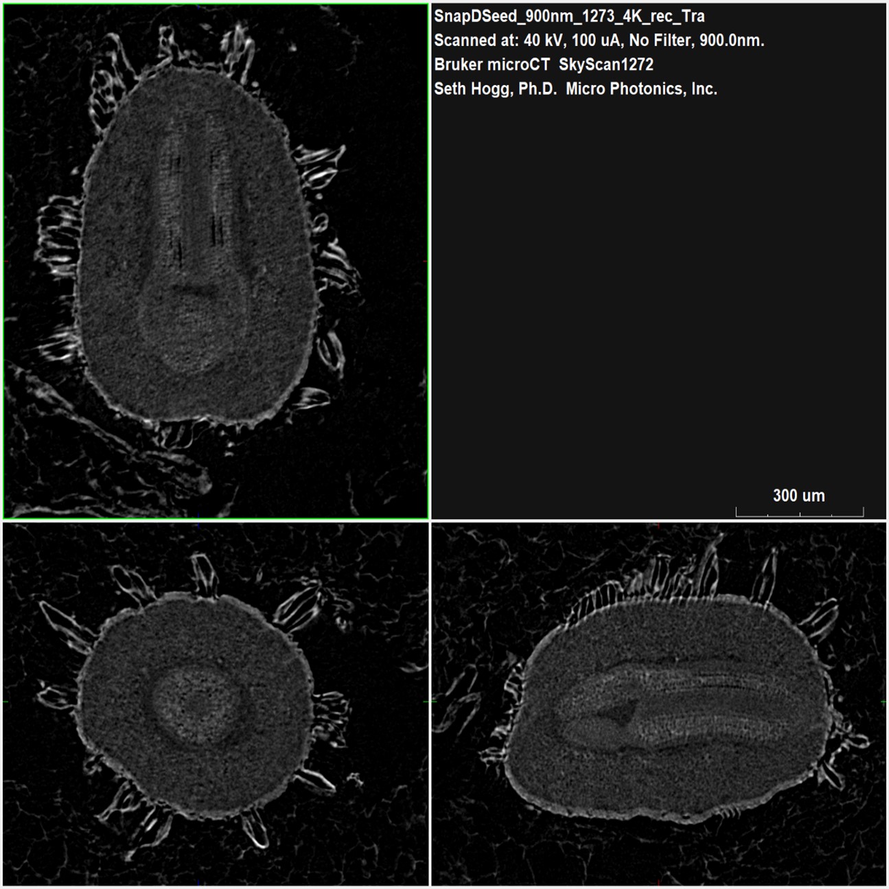
Figure 3: Planar 2D high-resolution views through an isolated seedMoving in from our overview imaging of the entire pod, Figure 3 highlights the level of detail captured of the internal and external features of the seed. While we were able to visually identify a seed in our pod dataset, this higher resolution view through the seed shows individual components of the seed from the dual phases central to its core and the dense outer shell with many projecting structures to both protect the seed and aid in attachment to a suitable carrier.
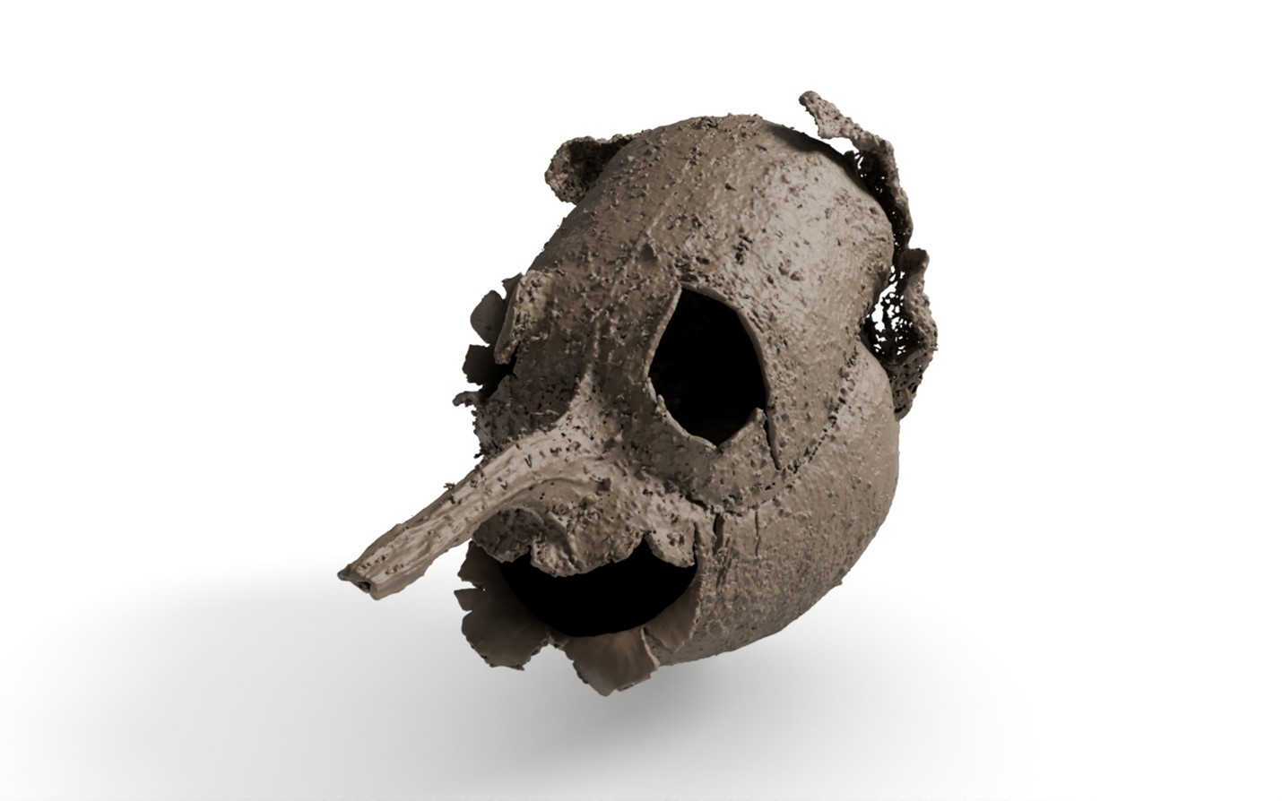
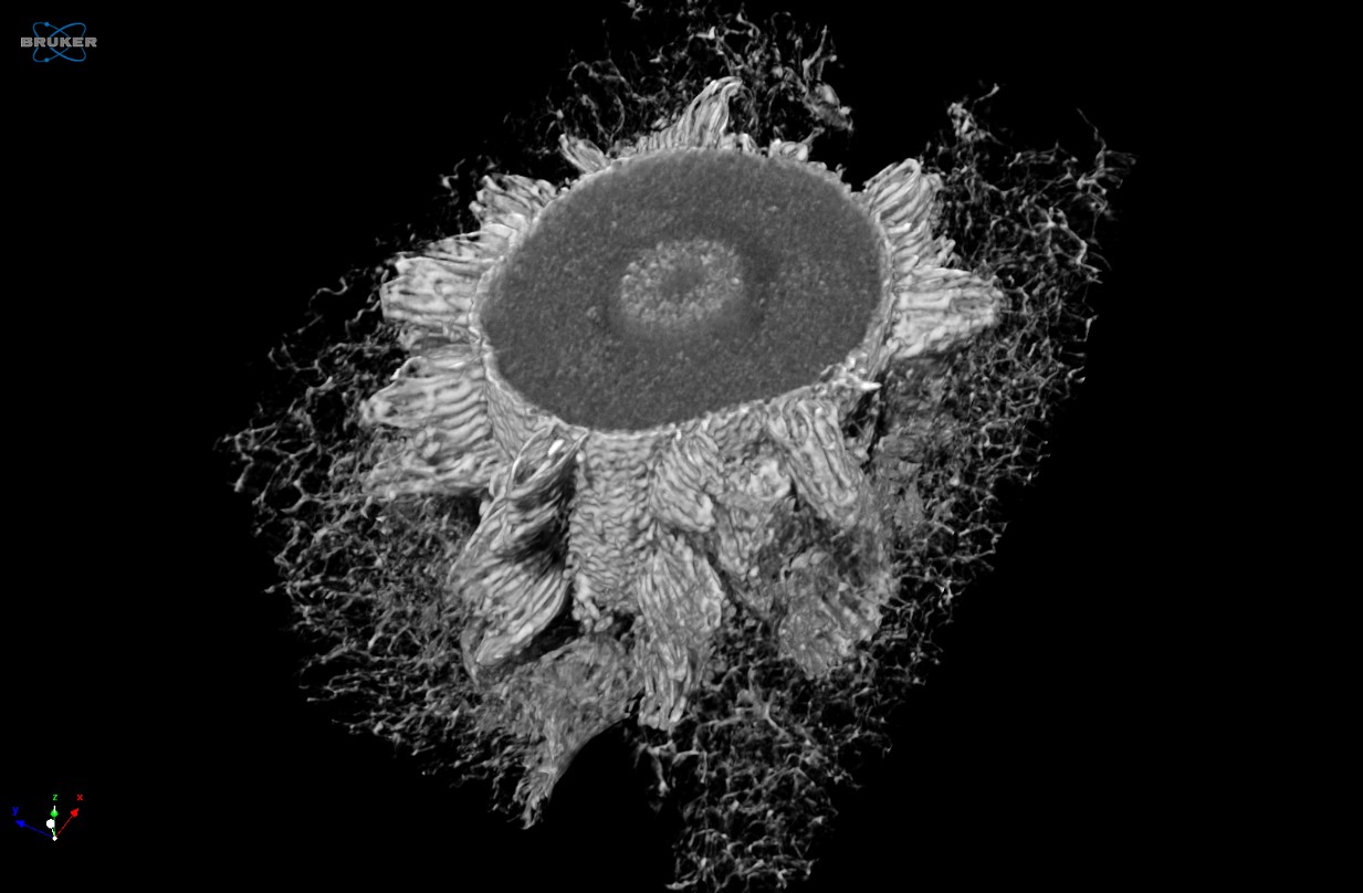
When we view the seed pod dataset in 3D with CTVox, we are greeted with an unusual surprise (Figure 4). The top portion of the seed pod bears an amusing resemblance to a face. The holes present in the pod to allow the seeds to leave after maturity are arranged in such a unique way.
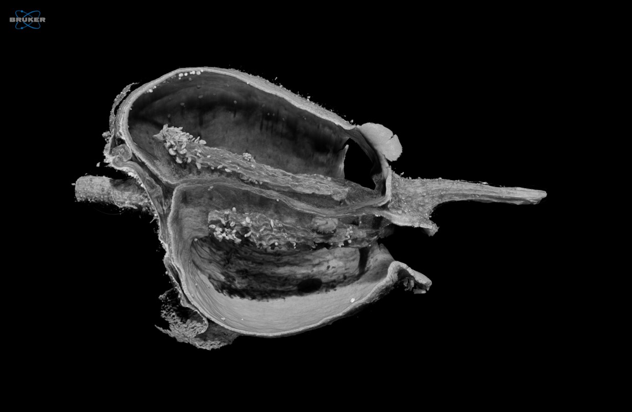
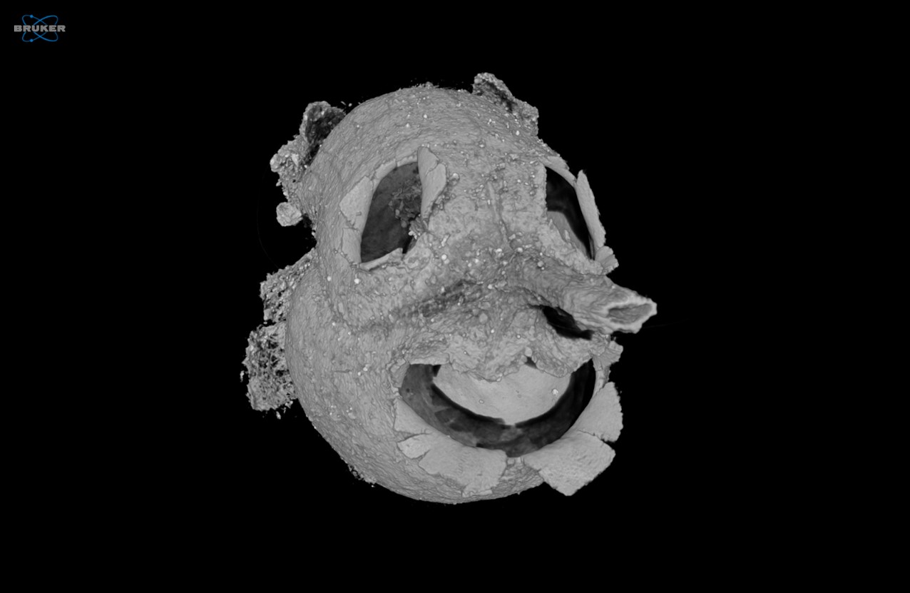
Using CTVox, we digitally sliced into the pod sample to reveal the inner structures, as shown in Figure 5. Much as we saw with the planar slices in DataViewer, the pod is primarily empty space at this point as presumably most of the mature seeds were already released from within the pod.
Similarly, we also digitally sliced into the seed dataset as shown in Figure 6, allowing us clear views of the seed coat, endosperm, and embryo. While the 2D view provided by DataViewer helped us understand the unique outer morphology of the seed coat, the full 3D rendering from CTVox provides an additional level of detail on the nature of these surface structures.
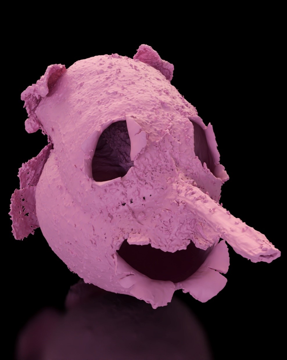
Figure 7: Colorized Maverick 3D rendering of the seed pod top surface
From our reconstructed SkyScan 1272 dataset, we imported the voxel-based seed pod dataset into Simpleware ScanIP software with the CAD add-on module to segment into a volumetric model. Maverick Render Indie then allowed us to apply colors and textures to the model, as shown in Figure 7.
Conclusion
Despite the launch and excitement around the new X4 Poseidon, the SkyScan 1272 remains an excellent micro-CT instrument for multiscale high-resolution imaging of organic samples like the pod and extracted seed imaged here.
We hope you found this Image of the Month informative and encourage you to subscribe to our newsletter and social media channels in preparation for the continuation of our Image of the Month series next month.
Scan Specifications
| Sample | Seed Pod | Single Seed |
| Voltage (kV) | 40 | 40 |
| Current (µA) | 150 | 100 |
| Filter | None | None |
| Voxel Size (µm) | 8 | 0.9 |
| Rotation Step | 0.3 | 0.25 |
| Exposure Time (ms) | 300 | 2097 |
| Rotation Extent (deg.) | 360 | 180 |
| Scan Time (HH:MM:SS) | 01:00:20 | 03:11:46 |
These scans were completed on our Bruker SkyScan 1272 micro-CT CMOS Edition at the Micro Photonics Imaging Laboratory in Allentown, PA. Reconstructions were completed using NRecon 2.0 while visualization and volumetric inspection of the 2D and 3D results were completed using DataViewer and CTVox. The seed pod dataset was converted to a STL volumetric model using Synopsys’ Simpleware ScanIP software with the CAD add-on module (Synopsys, Inc., Mountain View, USA) before 3D rendering using Maverick Render Indie (Random Control, Madrid, Spain).
Would you like your work to be featured in our monthly newsletter? If so, please contact us by calling Seth Hogg at 610-366-7103 or emailing seth.hogg@microphotonics.com.
References
*Simpleware software (Synopsys, Inc., Mountain View, USA) enables you to comprehensively process 3D image data (MRI, CT, micro-CT, FIB-SEM…) and export models suitable for CAD, CAE and 3D printing. Use Simpleware software’s capabilities to visualize, analyze, and quantify your data, and to export models for design and simulation workflows. Simpleware™ is a trademark of Synopsys, Inc. in the U.S. and/or other countries.
