
- Products
- Micro CT Systems
- SkyScan 1276
SkyScan 1276


The SkyScan 1276 is a high performance, stand-alone, fast, desktop in-vivo micro-CT with continuously variable magnification for scanning small laboratory animals (mice, rats, …) and biological samples. An optional Large Animal Transport System (LATS) is available which enables the system to mount also large animals including rabbits.
The SkyScan 1276 has an unrivalled combination of high resolution, big image size, round and spiral (helical) scanning and reconstruction, and low dose imaging. It administers a low radiation dose to the animals allowing multiple scans in longitudinal preclinical studies without the risk of unwanted radiation-induced side effects.
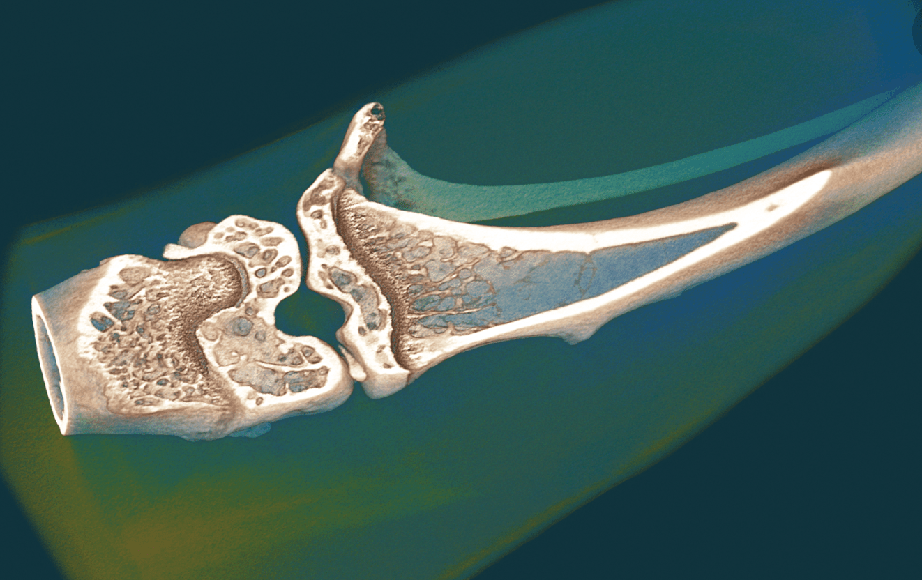
The variable X-ray energy combined with a range of filters ensures optimal image quality for diverse research applications from lung tissue to bone with metal implants.


Features
LARGE FORMAT DETECTOR
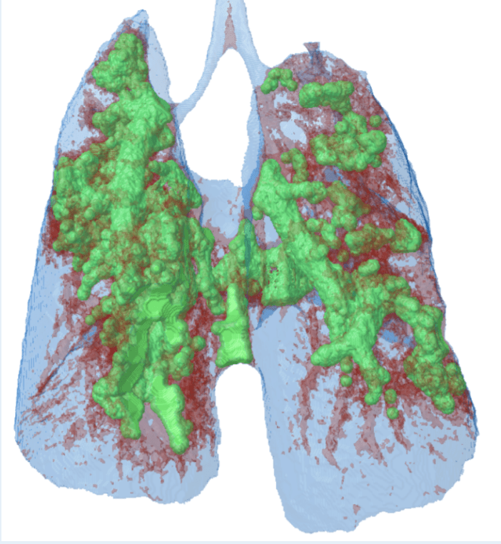
The large format array of the SkyScan 1276 allows for more volume to be acquired in a single scan rotation, reducing the overall dose by not having to scan multiple segments as well as the ability to capture full lung or heart images in synchronized gating acquisition mode.
VARIABLE MAGNIFICATION
Most in vivo micro-CTs on the market use a fixed working distance due to the fact that the source and detector must rotate around the specimen bed. In the SkyScan 1276, variable magnification is possible allowing for selection of the ideal resolution for in vivo scanning or making it possible to scan ex vivo at even higher resolution.
CONTINUOUS GANTRY ROTATION
The SkyScan 1276 can scan in continuous rotation mode to capture the length of the animal in unprecedented time.
ON-SCREEN REAL-TIME DOSE METER
The SkyScan 1276 control software includes a real-time on-screen dose meter. It indicates an estimation of the dose absorbed by the animal body during scanning. The measurement is based on the absorption calculated from X-ray projection images of the animal cross-calibrated with electronic dosimeter measurements. The dose meter shows accumulated dose or dose rate. It is calibrated for X-ray absorption in the standard mouse and rat cassettes. In this way it measures the X-ray dose absorbed in animal body itself during scanning. The dose absorbed by the animal during a scan is documented in the scan log-file together with all scan and reconstruction settings.
—–
SOFTWARE
The SkyScan 3D Software Suite is supplied as an unlimited site license so each user can have a copy; ensuring workflow from multiple groups is easily managed. Segmented software programs means post acquisition reconstruction, analysis, and modeling can be done on different computers, freeing the system for scanning. Image file outputs are standardized to TIF, BMP, or JPG formats for easy viewing, transferring, or copying.
Specifications
- X-ray Source
Continuously variable 20-100 kV micro-focus X-ray source
< 5 µm spot size at 4 W
Automatic 6-position filter changer - X-ray detector
16 Mp sCMOS detector (4096 x 4096 pixels) - 6-positions, automatic filter changer for energy selection
No filter, Al 0.2mm, Al 0.5mm, Al 1mm, Al 0.5mm + Cu 0.03mm, Cu 0.1mm (all filters > 99.9% purity) - Resolution
2.8 µm smallest pixel size
5-6 µm details resolved with more than 10% contrast - Maximum Object Size
75 mm diameter
310 mm height - Physiological Monitoring
Real-time motion detection (5 Mp color camera)
ECG, breathing detection, temperature stabilization
All signals digitized in 16bit with up to 120 samples/sec - Radiation Safety
< 1 µSv/h at any point on the instrument surface during scanning - Dimensions
954 mm (W) x 1190 mm (1560 mm with open door) (D) x 940 mm (H)
360kb (Weight) - Power Supply
100-240V AC, 50-60Hz, 3A max.
Do you want to see Micro-CT results of your sample?
Our specialists are happy to help.
Do you want to see Micro-CT results of your sample?
Our specialists are happy to help.
