
Micro-CT technology has transformed how zoologists conduct integrative and comparative biology research. Using 3D nondestructive imaging, brittle or complex biological samples can be fully analyzed without damaging the original sample. Micro-CT offers high sensitivity to different tissue types along with superior resolution for resolving subtle differences in morphometry. Our study of a preserved turtle specimen also allows us to showcase comprehensive visualization software and the stunning images that can be produced from micro-CT scans.
Among research into reptiles, the shells of turtles provide a notable point to study both in comparing existing species and historical ones. As dense bony structures, turtle shells are often well preserved during the fossilization process, providing a vast repository of structural information on these historical specimens. Micro-CT provides a non-destructive pathway towards preserving and sharing these structural details amongst the scientific community. Through imaging the samples, volumetric datasets are produced which can be freely shared worldwide.
X-Ray Microscopic Imaging of Reptiles
For this month’s imaging article, we examined a preserved turtle specimen using our high-powered SkyScan 1273 micro-CT at an isotropic voxel size of 45µm. The SkyScan 1273 is a great match for this project due to its high X-ray power (130kV at 300uA) as well as the large sample chamber.
Since our turtle specimen is approximately 7-8 inches in length and 5 inches in width, we utilized the full field of view of the SkyScan 1273 by activating the offset imaging mode. In this imaging mode, the X-ray detector offsets to each side of the sample chamber and a pair of acquisitions are completed to capture a wider horizontal field of view at a specified voxel size than would be possible with a traditional imaging mode. Finally, this offset imaging mode was applied at three points along the length of the sample to provide coverage on the full 7-8 inches, which resulted in a total volume comprised of six individual scans that are digitally combined into one dataset within NRecon.

As shown in Figure 2, DataViewer is used to straighten and align datasets after reconstruction. The ability to load datasets into a linked 3D viewing mode provides great control over the final orientation and cropping of your dataset. For the turtle sample imaged in this article, the sample was aligned horizontally in the coronal view to facilitate cropping the dataset into a smaller volume to save disk space and increase loading performance. The ability to fine tune the physical orientation of the dataset after imaging means that exact positioning of the sample in the instrument when setting up the acquisition is less important.

As shown in Figure 3, CTVox provides us with an interactive volumetric model of the dataset rendered in real time and often helps provide a clearer view of the location of features within the sample in 3D space compared to the representations possible in DataViewer. Using the histogram for the dataset, CTVox is able to isolate only the signal arising from the skeleton in the dataset while excluding the signal from the plastic container that housed the sample during imaging as well as the signal from the hydrated soft tissues present in the turtle’s abdomen.

Using clipping planes within CTVox, we digitally dissect the live view of the dataset as shown in Figure 4. In this view, we observe the vertebrae from the skull through the neck into the body of the turtle as well as clipped views of the pelvic and shoulder girdles. Some dense granules are also visible in the abdomen of the animal, perhaps the result of some undigested mineral contents from a leftover meal.

Within our laboratory, we also often utilize Synopsys Simpleware ScanIP software (U-2022.12-SP1; Synopsys, Inc., Mountain View, USA) to import our Bruker SkyScan micro-CT data and then produce optimized 3D models. As with CTAnalyzer, Simpleware ScanIP software allows us to utilize a traditional global threshold to select portions of our sample, which was suitable for this sample since we only wanted to isolate one component of the dataset, the skeleton (Figure 5).
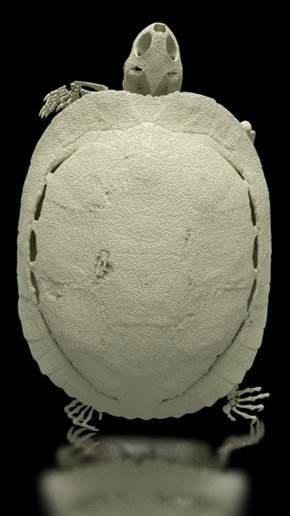
After isolating the skeletal volume from the larger dataset and creating a volumetric mesh, we’re free to move into any downstream 3D software suite to complete further work. In our case, we imported our mesh into Maverick Render Indie to create high resolution photo realistic renderings and videos of our scan data as shown in Figures 1 and 6, highlighting different views of the skeletal bones.
Conclusion
Among the SkyScan product line, the SkyScan 1273 stands alone for its versatility as it can accommodate the widest range of sample sizes to image samples from small to quite large (~10 inches). This versatility is of particular importance for researchers exploring zoological samples due to the vast disparities and size and composition between different projects that can be encountered. For this project, we worked near the maximum field of view of the SkyScan 1273 to capture a high-resolution dataset of the large turtle specimen. In combining both offset (double wide) and oversize (three vertical fields of view) imaging modes, the SkyScan 1273 captured fine details within the bones of our turtle sample despite the large physical size of the specimen.
We hope you found this Image of the Month informative and encourage you to subscribe to our newsletter and social media channels in preparation for the continuation of our Image of the Month series next month.
Scan Specifications
| Sample | Turtle |
| Voltage (kV) | 130 |
| Current (µA) | 300 |
| Filter | 2.0 mm Copper |
| Voxel Size (µm) | 45 |
| Rotation Step | 0.2 |
| Exposure Time (ms) | 800 |
| Rotation Extent (deg.) | 360 |
| Scan Time (HH:MM:SS) | 17:09:18 |
These scans were completed on our SkyScan 1273 micro-CT system at the Micro Photonics Imaging Laboratory in Allentown, PA using a combination of both offset and oversize imaging modes. Reconstructions were completed using NRecon 2.0 while visualization and volumetric inspection of the 2D and 3D results were completed using DataViewer and CTVox. The turtle skeleton was converted to a STL volumetric model using Synopsys’ Simpleware ScanIP software software with the CAD add-on module (Synopsys, Inc., Mountain View, USA) before 3D rendering using Maverick Render Indie (Random Control, Madrid, Spain).
Would you like your work to be featured in our monthly newsletter? If so, please contact us by calling Seth Hogg at 610-366-7103 or e-mailing seth.hogg@microphotonics.com.
References
https://en.wikipedia.org/wiki/Turtle_shell
*Simpleware software (Synopsys, Inc., Mountain View, USA) enables you to comprehensively process 3D image data (MRI, CT, micro-CT, FIB-SEM…) and export models suitable for CAD, CAE and 3D printing. Use Simpleware software’s capabilities to visualize, analyze, and quantify your data, and to export models for design and simulation workflows. Simpleware™ is a trademark of Synopsys, Inc. in the U.S. and/or other countries.
https://www.synopsys.com/simpleware.html