
Polymer materials are widely used as a basis for the manufacture of shoe soles and are superior in many respects to leather and other natural materials. The specific physical and mechanical properties of polymer sole materials vary greatly in characteristics such as elasticity, durability, frost-resistance, density, cushioning, strength, and wear resistance.
Micro-CT plays a key role in helping to investigate failure modes or defects in many types of polymers, nondestructively. X-ray microscopy is a powerful tool available to researchers and manufacturers to examine samples in their current state without the need for destructive testing.
X-Ray Microscopic Imaging of Defective Shoes
This month we imaged a pair of shoes with both a partial and a full failure of the polymer soles. We used the SkyScan 1273 micro-CT because of the large sample chamber volume, which allowed the shoes to easily fit without further modification. In failure analysis, it is key to minimize any changes to the product during the investigation and the ability to contain the full shoe without modification was central to this work. With the ability to image a sample up to both 10 inches in diameter and height, the SkyScan 1273 was a natural fit.


As shown in Figure 2, both shoes show a failure (cracking) in the soles in a similar location located under the ball of the foot. Since this region of the shoe experiences significant bending modes during normal wear, our first thought naturally leads to the idea of the failure occurring through dynamic fatigue of the sole.
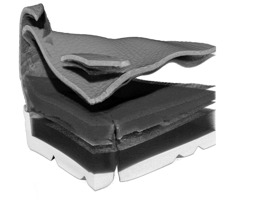
Exploring the data in 3D allows us to view the propagation of the crack through the sole into the interior layers of the shoe itself, including the insoles (Figure 3). The translation of the crack through different layers in the shoe lends credence to the theory regarding dynamic fatigue. If the source of the failure were due to a failure entirely related to the soles, we would not expect to see the structural effects throughout the layers of the shoe.
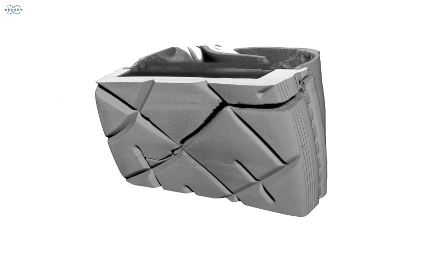
Examining the path of the crack shows us that the crack propagated along the natural pathways provided in the molding for the sole (Figure 4). As the shoe was continually flexed during walking and the cracks began to form at the base of the ball of the foot, it’s natural that the crack would propagate along the path of least resistance, and therefore cracking following the channels molded into the soles is expected.
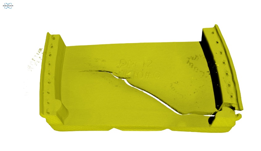
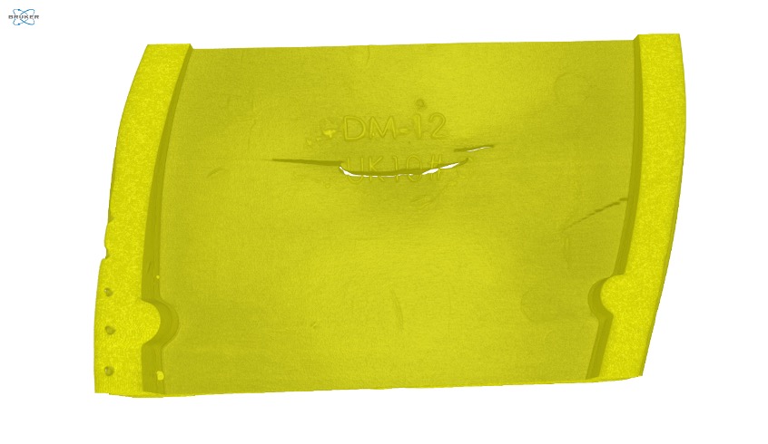
Micro-CT excels as a comparative technique, so having both a sample with a full failure of the sole and one with a partial failure provides us great insight into the sample. In this case, we see the failures for both shoes appearing in the region of embossed text molded into the soles denoting the brand and size of the shoes (Figure 5). Using CTVox, we isolated data only from the soles of the shoes to produce these images and allow a finer investigation of the soles without the signal from the rest of the shoe present. For both soles, the crack appears to have begun in a similar location and then propagated outward.
Conclusion
The large sample volume (imaging size up to 10×10 inches) of the SkyScan 1273 was a perfect match for the large shoe samples in this study. Rather than having to cut out and extract the failed portions of the shoes, each shoe was mounted in the instrument as received, without further modification and only the region containing the failed polymeric sole was imaged.
Based on the location and propagation of the cracks present in the failed polymeric properties, the results seem consistent with dynamic fatigue of the polymer. No large voids or other material defects were noted within the imaged portion of the polymeric sole.
We hope you found this Image of the Month informative and encourage you to subscribe to our newsletter and social media channels in preparation for the continuation of our image of the month series next month.
Scan Specifications
| Sample | Shoe Sections |
| Detector | Flat Panel |
| Voltage (kV) | 120 |
| Current (µA) | 300 |
| Pixel Size (µm) | 50 |
| Rotation Step | 0.2 |
| Scan Time (HH:MM:SS) | 02:34:47 |
These scans were completed on our SkyScan 1273 micro-CT system at the Micro Photonics Imaging Laboratory in Allentown, PA. Reconstructions were completed using NRecon while visualization and volumetric inspection of the 2D and 3D results were completed using Dataviewer and CTVox.
Would you like your work to be featured in our monthly newsletter? If so, please contact us by calling Seth Hogg at 610-366-7103 or e-mailing seth.hogg@microphotonics.com.