

Batteries are of growing importance from both a sustainability and energy storage standpoint as our world continues to require larger energy demands to sustain our lifestyles. Batteries are useful as a repository for excess energy produced during times when the supply exceeds the local demand and can be used, for instance, to store excess energy produced during the daytime with solar panels for release later at night when the solar panel production ceases. Batteries also provide a convenient, portable source of power for modern electronic devices.
Standard alkaline batteries are ubiquitous in the market and their design and construction are well noted. Micro-CT imaging allowed us to examine an alkaline AA battery with high resolution views of each key component.
X-Ray Microscopic Imaging of Batteries
Wen examined an AA battery sample, which had developed a leak over years of infrequent use, using our high-powered SkyScan 1273 micro-CT at an isotropic voxel size of 10µm.

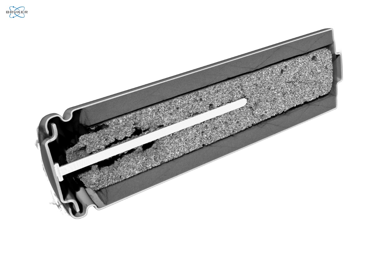
As shown in Figure 2, the SkyScan 1273 micro-CT provides a clear view through the AA battery and individual components of its construction are present. Notably, the potassium hydroxide (KOH) which leaked outside the battery to form the white deposits, is not visible in Figure 2 as the X-ray attenuation of the porous KOH film is closer to air than to the larger attenuation of the individual battery components. Notably, the negative cap, negative collection pin, outer metal can, manganese oxide cathode, and the zinc oxide cathode are all visible.
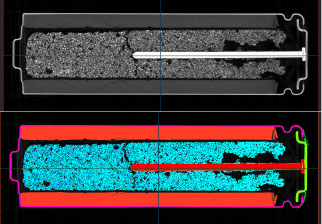
When moving out to a volumetric view of each dataset, the full structure of the individual components is more evident (Figure 3). In investigating a possible leak path for the KOH, the interactive 3D view provided by CTVox can be particularly useful.

Using clipping planes or shapes within CTVox, we can spotlight regions of interest such as the anode within our battery sample, as shown in Figure 4. While clipping planes are useful to slice through the dataset, the available clipping shapes can also be used to subtract or isolate specific regions of the dataset with more geometrical freedom than is possible with standard cubic faces.
The volumetric datasets provided by micro-CT imaging are ideal for conversion to 3D models, which can be used for computational simulations or additive manufacturing. One key step in the process is the segmentation of a grayscale dataset arising from micro-CT imaging into a distinct model. The most typical way segmentation occurs in Bruker CTAnalyzer is through thresholding, with global thresholding being the most popular. In many cases, different portions of the sample may be represented in the grayscale images with overlapping intensity values, requiring either careful definition of a region of interest or several cleanup steps to isolate only the feature of interest for modeling.
Within our laboratory, we also often utilize Simpleware ScanIP software (Version U-2022.12-SP1; Synopsys, Inc., Mountain View, USA) to import our Bruker SkyScan micro-CT data and then produce optimized 3D models. As with CTAnalyzer, Simpleware ScanIP software allows us to utilize a traditional global threshold to select portions of our sample. However, in this case a background flood fill tool is a better option to allow us to digitally isolate each component of the battery into a separate mesh which would be more challenging to try to complete within CTAnalyzer (Figure 5). With the background flood fill tool in Simpelware ScanIP software, you can define a seed starting point in your background images and select an upper and lower threshold value to be included or excluded from your selection. From this point, the software will grow the seed point in 3D space within the bounds of your threshold selection to capture the component of interest.

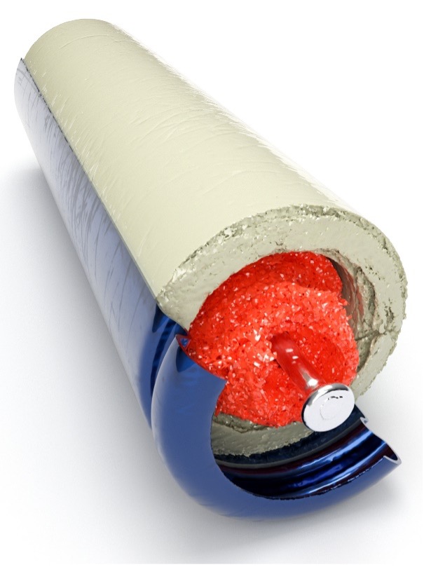
Once we’ve isolated our relevant features and created unique modeled meshes for each, we’re free to move into any downstream 3D software suite to complete further work. In our case, we imported our meshed into Maverick Render Indie to create high resolution photo realistic renderings and videos of our scan data as shown in Figure 6, highlighting both the negative cap of the battery as well as the anode collector pin.
Just like inspecting individual components, we can also inspect assemblies of subcomponents as a larger dataset to note relative position and assembly details. Once again, we used Maverick Render Indie to create a high-resolution photo realistic rendering of a clipped view through the metal battery can, highlighting the cathode as well as the anode and anode collector pin as shown in Figure 7.
Conclusion
Among the SkyScan product line, the SkyScan 1273 stands alone for its versatility as it can accommodate the widest range of sample sizes to image samples from small to quite large (~10 inches). For this project, we worked near the maximum resolution of the SkyScan 1273 to capture a high-resolution dataset of the defective AA battery sample.
We hope you found this Image of the Month informative and encourage you to subscribe to our newsletter and social media channels in preparation for the continuation of our Image of the Month series next month.
Scan Specification
| Sample | AA Battery |
| Voltage (kV) | 130 |
| Current (µA) | 115 |
| Filter | 2.0 mm Copper |
| Voxel Size (µm) | 10 |
| Rotation Step | 0.3 |
| Exposure Time (ms) | 800 |
| Rotation Extent (deg.) | 360 |
| Scan Time (HH:MM:SS) | 12:21:28 |
These scans were completed on our SkyScan 1273 micro-CT system at the Micro Photonics Imaging Laboratory in Allentown, PA. Reconstructions were completed using NRecon 2.0 while visualization and volumetric inspection of the 2D and 3D results were completed using DataViewer and CTVox. Individual components were meshed using Synopsys’ Simpleware ScanIP software software with the CAD add-on module (Synopsys, Inc., Mountain View, USA) before 3D rendering using Maverick Render Indie (Random Control, Madrid, Spain).
Would you like your work to be featured in our monthly newsletter? If so, please contact us by calling Seth Hogg at 610-366-7103 or e-mailing seth.hogg@microphotonics.com.
References
– https://www.betterbattery.co/blogs/blog/how-to-clean-and-dispose-of-corroded-batteries
– https://www.britannica.com/technology/battery-electronics/Primary-batteries
*Simpleware software (Synopsys, Inc., Mountain View, USA) enables you to comprehensively process 3D image data (MRI, CT, micro-CT, FIB-SEM…) and export models suitable for CAD, CAE and 3D printing. Use Simpleware software’s capabilities to visualize, analyze, and quantify your data, and to export models for design and simulation workflows. Simpleware™ is a trademark of Synopsys, Inc. in the U.S. and/or other countries.
