It is summer and is time to soak up some sun and head to the beach! As you walk along the beach you probably have collected seashells as a kid or still do. Those fond memories gave us the great idea to scan seashells.
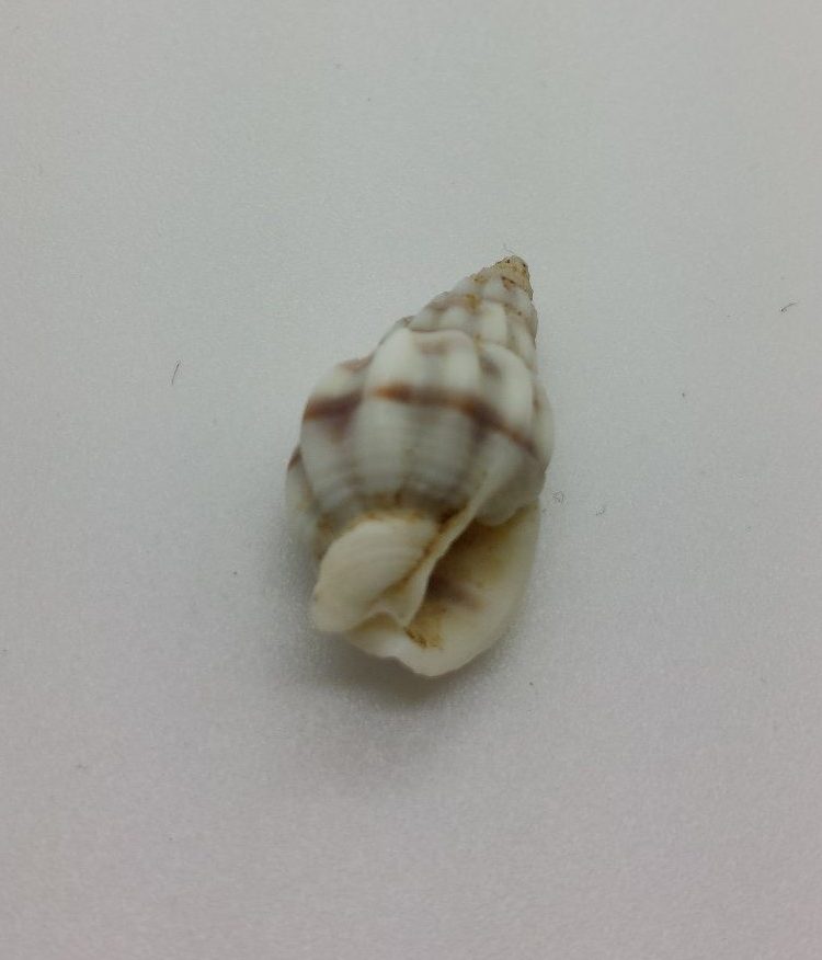
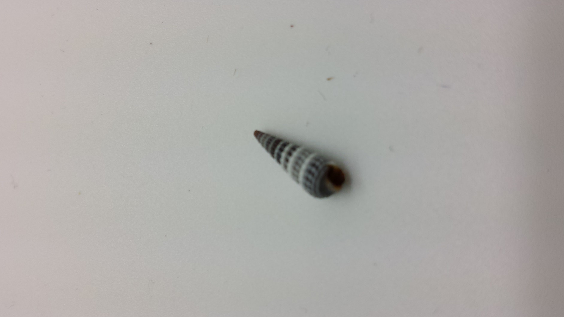
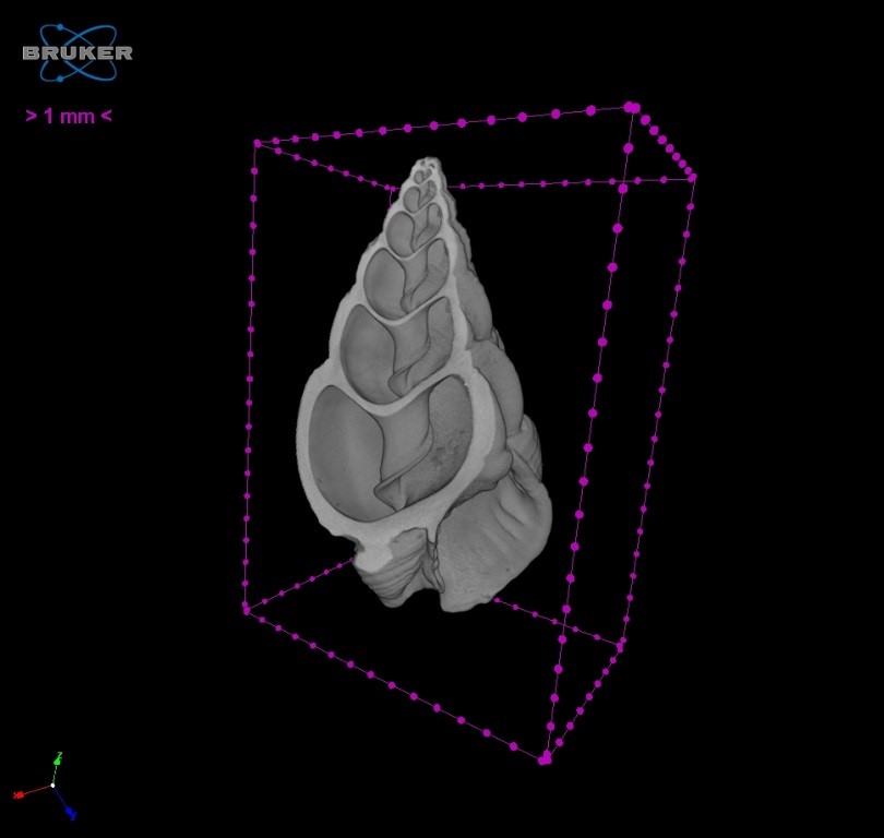
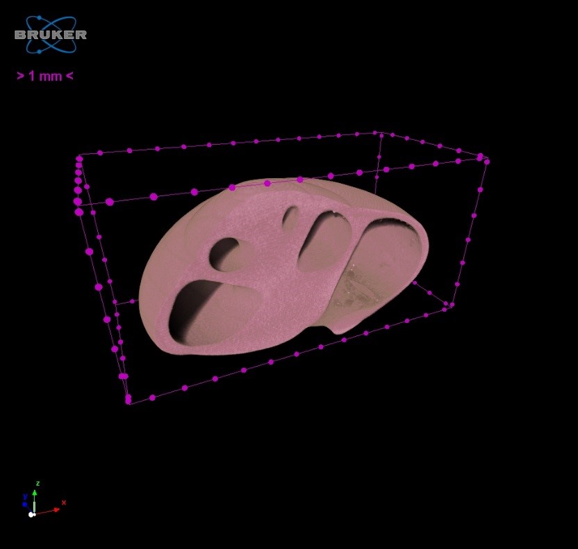
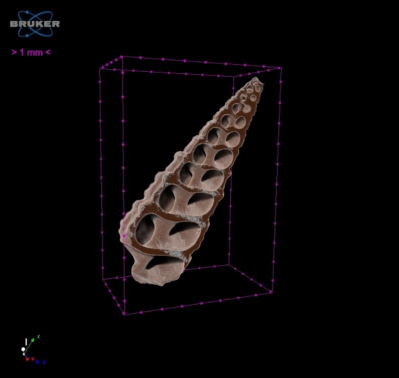
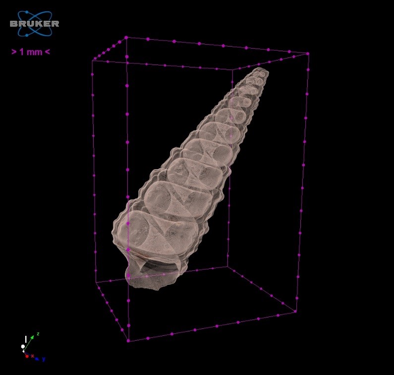
Combing the beach for seashells may turn up a variety of shapes, sizes and colors. As we sorted through the ones available to us, we could not just pick one so we ended up scanning three different varieties. They include a Nassa, Umbonium, and Auger seashells (See Figures 1, 2, and 3, respectively).
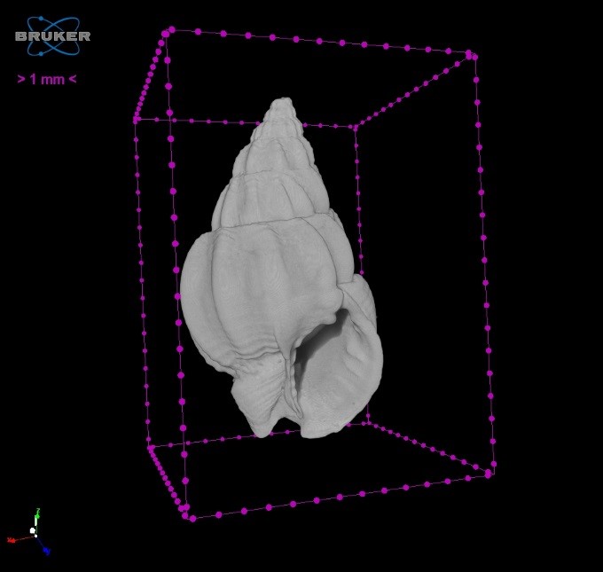

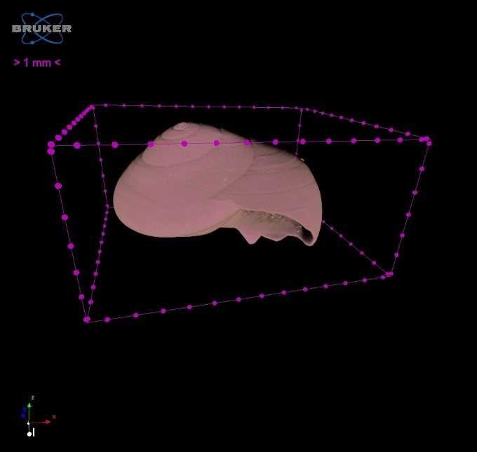
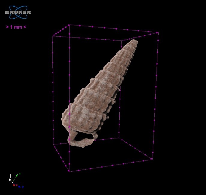
Using our Skyscan 1172 we were able to scan each of these seashells to capture their unique details and even provide stunning internal views (See Figures 4, 5, and 6, respectively). These internal structures of the shells are formed from the exoskeletons of mollusks, such as snails or oysters. They are mainly made up of calcium carbonate and some protein. As the mollusk grows the added calcium carbonate and protein are added to the fringes of the shells edge. Over time, these exquisite structure are formed.
References:
http://www.suzannesseashells.com/about-shells.php
http://www.scientificamerican.com/article/how-are-seashells-created/
System
SkyScan 1172 High Resolution Micro-CT
Voltage
65kV
Current
153µA
Pixel Size
Nassa – 13.37µm
Umbonium and Auger – 10.43µm
Rotation Step
Nassa – 0.3
Umbonium and Auger – 0.4
Scan Time
Nassa – 01:39:09
Umbonium and Auger – 00:46:28
Software
NRecon, DataViewer, CTvox
Location
Micro Photonics Imaging Laboratory, Allentown, PA
Courtesy of
Tim Sledz and Brandon Walters, Micro Photonics