Micro-CT for Entomology
Can the built environment mimic the natural world? Biomimicry — analyzing structures in nature in order to understand how they function — allows engineers to bridge the gap between designers and nature but requires a deep knowledge of the structures being analyzed. One way to learn more about these structures is to examine them via micro-CT imaging. Such imaging provides a path towards generating a digital model of any feature, which can be further explored via computer simulation or additive manufacturing.
Many researchers focus on the development of new technologies based on inspiration derived from the study of insects. Studying wings, eyes, and other insect structures provides opportunities for new technological advances using biomimicry and reverse engineering.123 For our study of micro-CT for entomology we utilized our high resolution SkyScan 1272 scanner to look at a common house fly (Musca Domestica). We chose to use micro-CT to visualize the entire insect rather than focusing on a single feature, such as the head or wings.
Micro-CT Scan of a House Fly
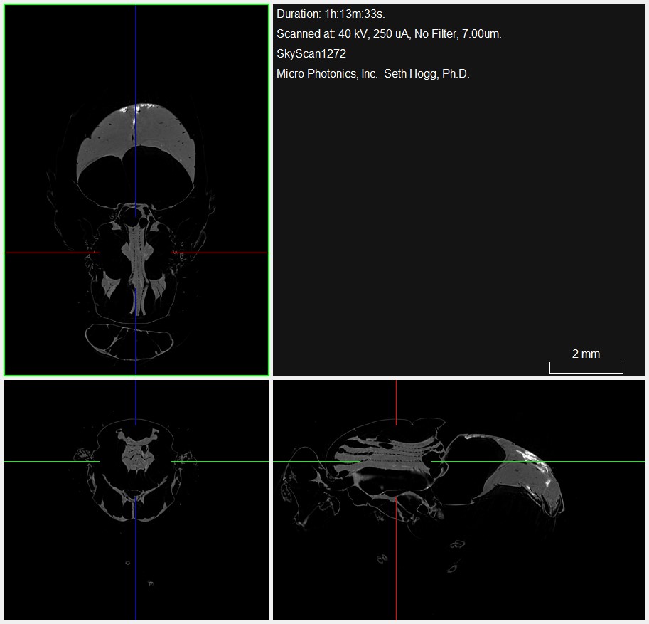
Utilizing the reconstructed X-ray attenuation data, two-dimensional views through the fly were generated (Figure 1). These views allow us to examine the head, thorax, and abdomen of the insect and visualize the associated internal structures.

The reconstructed images also provide a full three-dimensional rendering of the fly, which can be positioned and sliced as needed to provide greater dimensional context to the internal features (Figure 2).


For a closer inspection of features of interest, regions of the sample can be isolated into volumes of interest for further examination (Figure 3). In this case, the head of the fly is isolated to view without the data from the rest of the insect, allowing us to more closely visualize this feature.
Conclusion
Micro-CT lets us peek inside the body to learn more about the anatomy and structure of insects. However, this ability is not limited to insect analysis and is also useful in fields ranging from structural biology and material science to geology, odontology, and many others. We hope you found this Image of the Month informative. If you have an Image of the Month sample that you would like us to scan, please contact us by calling Seth Hogg at 610-366-7103 or e-mailing seth.hogg@microphotonics.com
Scan Specifications
| Sample | House Fly (Musca Domestica) |
| Voltage (kV) | 40 |
| Current (µA) | 250 |
| Pixel Size (µm) | 7 |
| Rotation Step | 0.2 |
| Scan Time (HH:MM:SS) | 01:13:33 |
This scan was completed on our high energy SkyScan 1272 micro-CT system at the Micro Photonics Imaging Laboratory in Allentown, PA. Reconstructions were completed using NRecon and visualization of 2D and 3D results were completed using DataViewer and CTVox.
Works Cited