Product Development: Using Micro-CT to Examine Medical Devices

The manufacturing of medical devices involves long processes that face global competition. Requirements for the highest levels of quality, reliability, and safety continue to rise as new technologies are evolving. Micro-CT allows for a deep investigation of products through nondestructive testing (NDT) and is a tool that contributes to quality improvement all along the supply chain through verifying integrity, measurements, and the detection of defects.
Examination of an EpiPen by micro-CT
In this micro-CT demonstration, we examined a single use medical device, an EpiPen, with a complicated construction. In this EpiPen, a syringe is in a retracted position within a plastic shell containing a dose of epinephrine. When in operation the syringe moves downward, causing the needle to protrude for penetration into the thigh of the patient. The medication is subsequently dispensed through a spring application of pressure on the syringe plunger. Micro-CT allows us to nondestructively examine the relationship of the parts to one another and to visualize possible defects that occurred during the manufacturing process.
For this month’s demonstration, we chose to utilize our Bruker SkyScan 1173 system because of its capacity to handle larger size samples; the 1173 is capable of high spatial resolution with samples that have a high aspect ratio, such as the device examined in this study.

Micro-CT Scan of a Medical Device
After reconstructing the X-ray attenuation data for the medical device, we were able to isolate our view on specific regions of interest. One such location is the interface between the syringe plunger and the syringe body (Figure 1). In this device, the syringe needs to actuate quickly and reliably in the event of an emergency medical crisis. Other important areas of inspection would include the springs located both at the front and rear of the device that are also of critical importance during use.
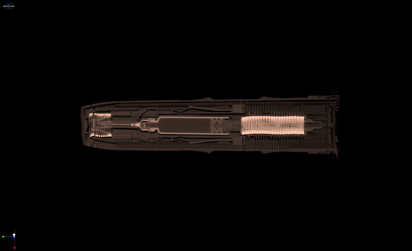
Taking a look from a side view in a three-dimensional rendering, we can inspect the interaction of the components in a live view with depth (Figure 2). In the current closed state, the syringe plunger is still positioned at the entrance to the syringe barrel and the springs are all tightly compacted.
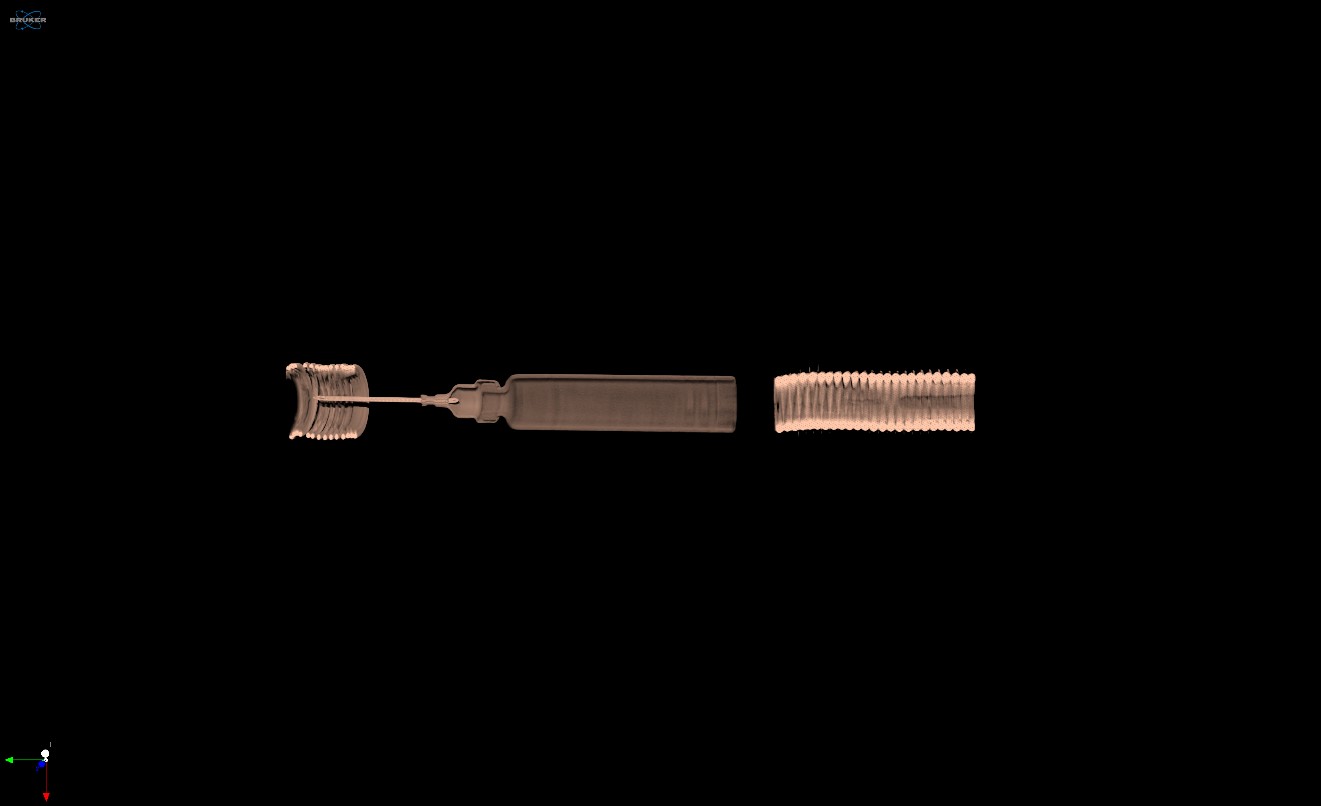
Using image filtering within our CTVox software, we can remove the low density plastic components from the view in Figure 2, allowing us to better visualize the dense metal and glass components comprised of the springs, needle, and syringe body (Figure 3).
Conclusion
Micro-CT excels as a tool to nondestructively examine medical devices and produces a detailed three-dimensional model that can be reoriented in any direction for examination. We hope you found this image of the month interesting. If you have an image of the month sample that you would like us to scan, please contact us by calling Seth Hogg at 610-366-7103 or emailing seth.hogg@microphotonics.com
Scan Specifications
| Sample | Medical Device |
| Voltage (kV) | 130 |
| Current (µA) | 61 |
| Filter | 0.25mm Brass |
| Pixel Size (µm) | 70 |
| Rotation Step | 0.5 |
| Scan Time (HH:MM:SS) | 01:28:45 |
All scans completed on our large capacity SkyScan 1173 micro-CT system at the Micro Photonics Imaging Laboratory in Allentown, PA. Reconstructions were completed using NRecon and visualization of 2D and 3D results were completed using DataViewer and CTVox.