Fiber-reinforced plastics (or polymers) are composite materials made of a polymer matrix that is reinforced with fibers. Typically, most of these composites have strong, stiff fibers in a matrix, which produces a component that often has a low density yet is strong and stiff. Fibers, such as glass (as in fiberglass), carbon, aramid, and basalt, are combined with epoxy, vinyl ester, or polyester thermosetting plastic to form the composite. These composites are used in numerous industries, including automotive, marine, aerospace, and construction. Micro-CT is ideal for analyzing individual fiber properties, such as the diameter, as well as the fiber distribution, and orientation.
We used our high resolution SkyScan 1272 desktop micro-CT scanner to examine selected regions of a one-piece composite hockey stick, demonstrating how micro-CT can be used for fiber characterization and orientation analysis of fiber-reinforced plastics.
Micro-CT Imaging of a Hockey Stick
To begin this study, we first examined a portion of the shaft removed from the stick. As can be seen in Figure 1 above, the full width of the stick could be accommodated at high resolution using a two-part offset scan. In this scanning mode, each horizontal half of the stick is scanned independently and then the two scans are digitally stitched together to form one extended-width image.
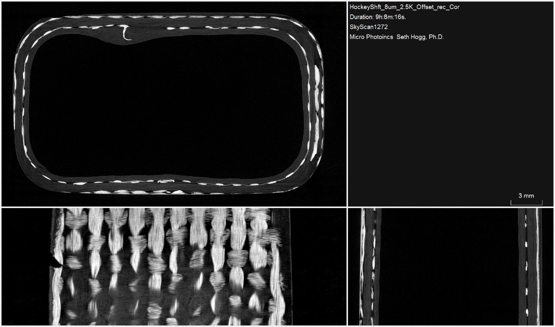
As can be seen in Figure 2, the manufacturer of the hockey stick incorporated a woven fiber support fabric into the polymeric extrusion to construct the shaft. The incorporation of the fabric into the shaft increases the strength and durability of the polymer to accommodate the stresses involved in the sport.
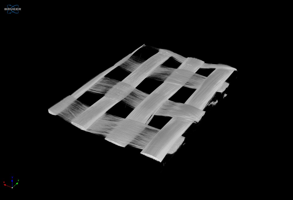
While we were generally able to resolve the fabric support layer from within the polymer, the individual fibers are not resolved from one another in the original offset scan. Further segmenting of the sample allowed us to scan a smaller region at increased resolution to find better definition among the fibers. Figure 3 highlights the fiber bundles incorporated into the fabric support structure from the scan data obtained at a voxel size of 2.25µm.
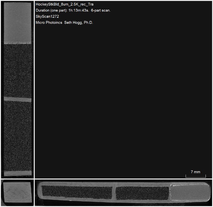
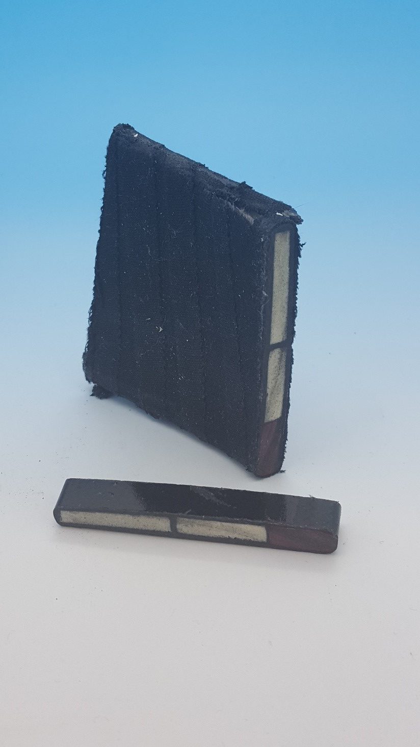
While the hockey stick shaft needs to be durable and flexible to create torque when shooting hockey pucks, the blade needs to be stiff while maintaining durability. To create the stiffness needed to facilitate the transfer of energy from the stick to the hockey puck, the manufacturer incorporated three chambers, each filled with a porous foam. As can be seen in Figure 4, when looking at the face of the hockey stick, the two top chambers are full of a low-density foam. This was indicated by the faint signal we observed after reconstructing the dataset. Meanwhile, the lowest chamber, which is in contact with the rough ice surface, is composed of a much higher density foam, as indicated by the increased image intensity in this volume.
Conclusion
Micro-CT provides information about sample composition over a wide range of feature sizes. While we can obtain useful information at larger voxel sizes, we can also image a smaller portion of a sample at higher resolution to better resolve fine structures, such as the fiberglass fibers comprising the support mesh incorporated in the hockey stick shaft. We hope you found this Image of the Month informative. If you have an Image of the Month sample that you would like us to scan, please contact us by calling Seth Hogg at 610-366-7103 or e-mailing seth.hogg@microphotonics.com
Scan Specifications
|
Sample |
Shaft Overview |
Shaft VOI |
Blade |
|
Voltage (kV) |
80 |
60 |
60 |
|
Current (µA) |
125 |
166 |
166 |
|
Pixel Size (µm) |
8 |
2.25 |
8 |
|
Rotation Step |
0.2 |
0.1 |
0.25 |
|
Scan Time (HH:MM:SS) |
09:08:16 |
04:38:44 |
01:13:43 |
This scan was completed on our high resolution desktop SkyScan 1272 system at the Micro Photonics Imaging Laboratory in Allentown, PA. Reconstructions were completed using NRecon and visualization of 2D and 3D results were completed using CTVox.