Automatic analysis of the 3-D microstructure of fruit parenchyma tissue using X-ray micro-CT explains differences in aeration
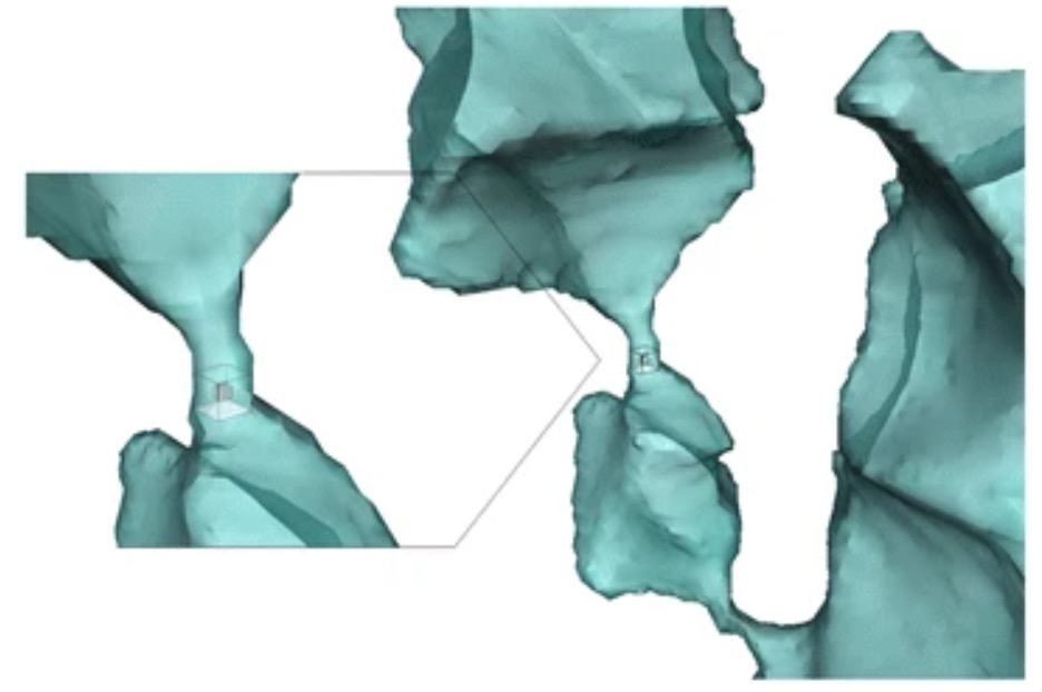
This study presents an automated method for processing high-resolution micro-CT images of parenchyma tissues of apple (Malus × domestica Borkh.) and pear (Pyrus communis L.) tissue, providing a quick objective method for characterizing 3D plant tissue anatomy at the level of single cells and intercellular spaces. “This will significantly aid interpretation and analysis of future tissue aeration studies. The automated image analysis methodology will also support pheno- and genotyping studies where the 3D tissue anatomy plays a role.”
…………………………………………………………………………………………………………………
X-ray computed tomography for 3D plant imaging
Plant researchers are increasingly using high resolution X-ray microscopy for structural phenotyping experiments at micro- and nanoscale levels. Automatic image-processing algorithms are now replacing manually operated segmentation procedures, and new developments in contrast agents and phase contrast are allowing the visualization of previously unresolved structural features. Mathematical models help compute how plant tissue structures affect relevant physiological processes such as respiration and photosynthesis. “Time-resolved X-ray CT techniques are emerging and permit dynamic imaging in real time owing to improvements in acquisition and image reconstruction.”
…………………………………………………………………………………………………………………
Probing the 3D architecture of the plant nucleus with microscopy approaches: challenges and solutions
Micro-CT and nanotechnology help researchers to more comprehensively resolve nuclear architecture in plants, which is a challenge as plant nuclei require novel solutions for capturing cell-specific, sub-nuclear, and dynamic processes. The eukaryotic cell nucleus is a central organelle and its architecture determines genome function at multiple levels. Utilizing high resolution X-ray microscopy as a tool, researchers are better able to deliver a comprehensive, 3D view of plant nuclear architecture and are also able to capture spatial dynamics of the nucleus relative to cellular states and responses.
………………………………………………………………………………………………………………
X-ray micro-computed tomography (micro-CT) of pyrite-permineralized fruits and seeds from the London Clay Formation (Ypresian) conserved in silicone oil: a critical evaluation
“Pyrite-permineralized fruits and seeds from the London Clay Formation (Ypresian; England) in the NHMUK are stored in silicone oil to retard decay processes. X-ray micro-computed tomography (micro-CT) has revealed internal morphology for multiple holotypes (including severely cracked and encrusted specimens) scanned in the protective fluid.” Iron pyrite permineralized fossils decompose quickly and a specimen can be totally destroyed when exposed to oxygen and high humidity. The plant fossils from the London Clay Formation in NHMUK are currently stored in silicone oil to limit or prevent these problems. Temporary removal of silicone oil from the surface of specimens for macrophotography or light microscopy requires washing in toluene, and places specimens at risk, so the ability to study the specimens with micro-CT without removing the protective silicone oil is a great benefit and protects the specimens for future study.
………………………………………………………………………………………………………………
Related Articles:
X-ray Microscopic Examination of An Apple
Micro-CT Imaging of a Strawberry Seed
Micro Photonics Inc. provides instruments, laboratory services, training, and support from micro-CT experts
to help research scientists meet their most complex laboratory demands.
More information: FREE BUYERS’ GUIDE
Request: FREE EVALUATION SCAN to see how micro-CT works with your application.
Contact: Benjamin Ache, Product Manager, Bruker Micro-CTs P: 610-366-7103 ext 115.